
Anju Batra Dr
About Me
Anju Batra Dr
DO
Ophthalmologist
10 Years Experience
Biography
My Clinic
OPD Hours
| Monday | Morning | 10:00 AM | - | 02:00 PM |
| Evening | 06:00 PM | - | 09:00 PM | |
| Tuesday | Morning | 10:00 AM | - | 02:00 PM |
| Evening | 06:00 PM | - | 09:00 PM | |
| Wednesday | Morning | 10:00 AM | - | 02:00 PM |
| Evening | 06:00 PM | - | 09:00 PM | |
| Thursday | Morning | 10:00 AM | - | 02:00 PM |
| Evening | 06:00 PM | - | 09:00 PM | |
| Friday | Morning | 10:00 AM | - | 02:00 PM |
| Evening | 06:00 PM | - | 09:00 PM | |
| Saturday | Morning | 10:00 AM | - | 02:00 PM |
| Evening | 06:00 PM | - | 09:00 PM | |
| Sunday | Morning | 10:00 AM | - | 02:00 PM |
| Evening | 06:00 PM | - | 09:00 PM |
Treatments
Specialised care and treatments offered
RETINAL LASER PHOTOCOAGULATION
This procedure utilizes targeted laser energy applied precisely to retinal areas affected by disease. It stabilizes conditions like diabetic retinopathy reducing risk of severe vision loss caused by abnormal blood vessels and leakage.
CORNEAL TRANSPLANTATION
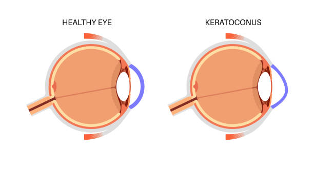
The cornea’s transparency is vital for clear vision; damage due to injury, infection, or dystrophies can necessitate transplantation to restore sight. This surgical replacement uses donor tissue for optimal outcomes.
VITRECTOMY
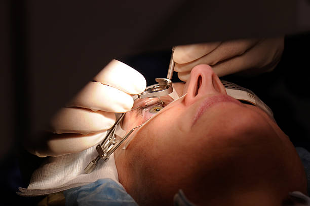
A vitrectomy is a microsurgical procedure performed to remove the vitreous gel from inside the eye. It treats various retinal disorders that impair vision by allowing access for repair or clearing opaque media obstructing sight.
- Candidates & Symptoms: Conditions like retinal detachment, diabetic retinopathy complications, or vitreous hemorrhage indicate need for vitrectomy surgery.
- Anatomical Assessment: Detailed retinal imaging guides surgical planning before intervention. Surgical Technique:The vitreous gel is carefully extracted through tiny incisions under high magnification while preserving retina integrity;
GLAUCOMA MANAGEMENT AND SURGERY
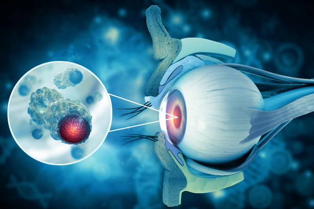
Glaucoma involves increased pressure inside the eye that can damage the optic nerve, potentially resulting in irreversible blindness if untreated. Early intervention is key to preserving vision.
- Disease Mechanism & Signs: Elevated intraocular pressure leads to gradual peripheral vision loss often without early symptoms.
- Detection: Measurement of eye pressure, optic nerve assessment via imaging, and visual field exams help confirm diagnosis.
- Treatment Options: Medical therapy includes pressure-lowering eye drops; surgical options like trabeculectomy or minimally invasive glaucoma surgeries (MIGS) reduce fluid buildup when needed.
- Surgical Expectations: Procedures aim to protect remaining nerve tissue; recovery times vary based on technique used.
Lifelong monitoring combined with timely surgery or medication management preserves eyesight in most glaucoma patients.
LASER-ASSISTED REFRACTIVE SURGERY (LASIK)
LASIK reshapes the cornea using laser technology to correct common vision problems such as nearsightedness, farsightedness, and astigmatism. It offers a minimally invasive alternative to glasses or contact lenses.
- Indications & Symptoms: Difficulty focusing on distant or close objects can prompt evaluation for refractive surgery.
- Evaluation: Thorough preoperative testing determines corneal thickness and suitability for LASIK.
- Surgical Process: A precision laser creates a corneal flap allowing reshaping of underlying tissue to improve focus.
- Postoperative Care: Patients often notice clearer vision within hours; mild dryness or irritation is common initially.
- Outcomes: Most achieve significant reduction in dependency on corrective lenses with minimal complications.
This surgery provides lasting visual clarity for many individuals seeking freedom from corrective eyewear.
CATARACT SURGERY
Cataracts cause clouding of the eye's natural lens, leading to blurry vision and difficulty seeing clearly. Surgery is the only effective solution to restore sight once cataracts interfere significantly with daily activities.
- Causes & Symptoms: Gradual vision loss, glare, faded colors, and difficulty at night are typical signs.
- Diagnosis: Comprehensive eye exams including slit-lamp and visual acuity tests identify cataract severity.
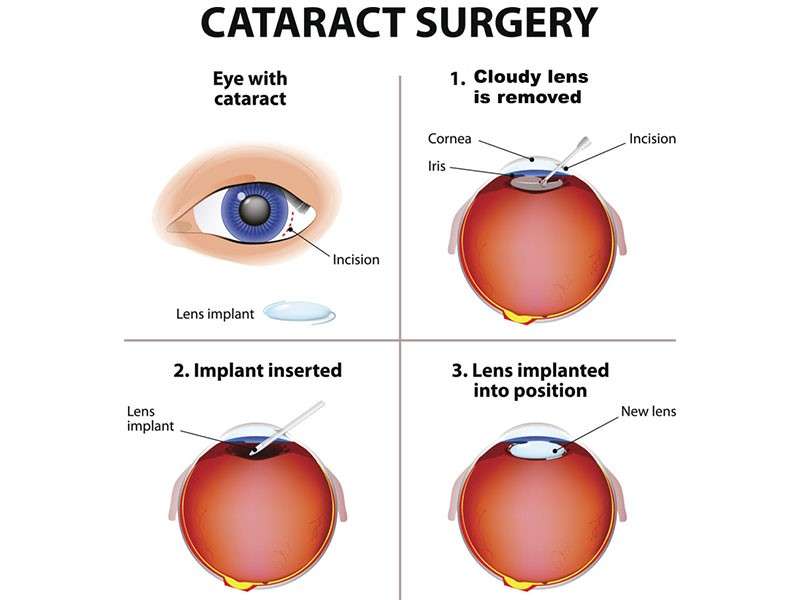
- Treatment: The cloudy lens is removed and replaced with a clear artificial intraocular lens (IOL) using advanced microsurgical techniques.
- Recovery: Most patients experience rapid improvement in vision; minor discomfort may occur temporarily.
- Follow-up: Regular check-ups ensure proper healing and optimal lens position.
This procedure effectively restores focus and contrast sensitivity, greatly enhancing quality of life after successful treatment.