
Dr Abhay Singh Ja
About Me
Dr Abhay Singh Ja
MS
Orthopaedic Surgeons
10 Years Experience
Biography
My Clinic
OPD Hours
| Monday | Morning | 10:00 AM | - | 02:00 PM |
| Evening | 06:00 PM | - | 09:00 PM | |
| Tuesday | Morning | 10:00 AM | - | 02:00 PM |
| Evening | 06:00 PM | - | 09:00 PM | |
| Wednesday | Morning | 10:00 AM | - | 02:00 PM |
| Evening | 06:00 PM | - | 09:00 PM | |
| Thursday | Morning | 10:00 AM | - | 02:00 PM |
| Evening | 06:00 PM | - | 09:00 PM | |
| Friday | Morning | 10:00 AM | - | 02:00 PM |
| Evening | 06:00 PM | - | 09:00 PM | |
| Saturday | Morning | 10:00 AM | - | 02:00 PM |
| Evening | 06:00 PM | - | 09:00 PM | |
| Sunday | Morning | 10:00 AM | - | 02:00 PM |
| Evening | 06:00 PM | - | 09:00 PM |
Treatments
Specialised care and treatments offered
BONE DENSITY TEST
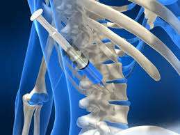
A bone density test measures how much calcium and other minerals are present within your bones. This test helps assess the strength of bones—critical in determining osteoporosis risk levels among older adults or those prone due to hormonal changes over time or medication use affecting skeletal health.
- Identifying Risk Factors: Often recommended following low-impact fractures indicating weakened bones people meeting age criteria should consider proactive assessment starting basis preventing future complications arising from fragile skeletal structures becoming more susceptible among demographic groups surveyed closely regarding association existing disease risks applicable them overall health profile reflected there.'
The importance underlined here shows preventative measures taken early could minimize incidence rates linked primarily brittle aspects found throughout framework we depend upon daily functionality wise.'
CT SCAN FOR BONE TUMORS

A CT scan (Computed Tomography) serves as an advanced imaging technique that allows doctors to visualize bones in cross-section. It plays an essential role in diagnosing bone tumors by providing clear images that assist in evaluating potential growths accurately.
- When Is It Needed? Patients presenting with persistent bone pain or unexplained swelling may be referred for this test if there's suspicion of tumor involvement based on prior exams/imaging results.
- How It's Conducted: The process involves lying on a table that moves through a donut-shaped machine while multiple X-ray images are taken around the area being examined producing detailed sections layered together into 3D format later analyzed by specialists.
- Analysis & Interpretation: Radiologists interpret images identifying potentially cancerous lesions among benign changes aiding physicians decide further management strategies needed post-diagnosis depending upon their findings.
- Treatment Considerations: If tumors are identified; discussions surrounding surgical removal vs alternative therapies begin ensuring comprehensive care plan established tailored to individual circumstances.
A CT scan represents one vital step towards accurate diagnosis allowing appropriate intervention based upon distinct findings made during evaluations.
X-RAYS FOR FRACTURES

X-rays are one of the primary imaging techniques used for diagnosing bone fractures. This quick and non-invasive method provides essential information regarding bone integrity when trauma occurs.
- Main Uses: X-rays are routinely employed after falls or accidents where fractures are suspected based on visible swelling or deformity in limbs.
The ability to quickly identify fractures aids in timely management which can lead to better outcomes during recovery periods.
SHOULDER MRI

An MRI (Magnetic Resonance Imaging) of the shoulder is a crucial diagnostic tool that provides detailed images of soft tissues, ligaments, tendons, and cartilage. It is instrumental in diagnosing various shoulder injuries or conditions that affect mobility and cause pain.
- Reasons for MRI: Common indications for an MRI include unresolved shoulder pain, rotator cuff tears, impingement syndrome, or suspected labral tears.
- How It Works: During the MRI scan, patients lie on a table that slides into a cylindrical machine where powerful magnets create detailed images without radiation exposure.
- Interpretation of Results: A radiologist analyzes the images to identify any abnormalities that could explain symptoms reported by the patient.
- Treatment Following Diagnosis: Depending on findings, treatment options may vary from conservative management like physiotherapy to surgical interventions if necessary.
An MRI is vital for your doctor in creating an informed treatment plan tailored to your specific needs.
HIP REPLACEMENT

Hip replacement surgery is a common but major procedure aimed at alleviating severe hip pain caused by arthritis or injury. The surgery entails removing damaged bone and cartilage from the hip joint and replacing it with artificial components to restore function.
- Symptoms Indicating Surgery: Persistent pain, stiffness in the hip joint, and difficulty performing everyday activities are key signs that may lead to this surgical intervention.
- Diagnosis: An orthopedic surgeon will perform a thorough evaluation including physical exams and imaging studies like X-rays to assess joint damage.
- Surgical Procedure: The procedure can be performed using traditional incisions or minimally invasive techniques depending on patient needs.
- Expectations Post-Surgery: Patients typically undergo rehabilitation involving exercises to improve mobility; most individuals report improved quality of life after recovery.
A hip replacement can significantly enhance functionality and decrease discomfort for individuals suffering from debilitating conditions.
KNEE ARTHROSCOPY

Knee arthroscopy is a minimally invasive surgical procedure used to diagnose and treat problems within the knee joint. This technique involves using a small camera known as an arthroscope, which allows the surgeon to visualize the internal structures of the knee without making large incisions.
- Common Symptoms: Patients often experience pain, swelling, stiffness, and a reduced range of motion in the knee.
- Diagnostic Approach: Diagnosis is typically achieved through physical examinations and imaging tests such as X-rays or MRI scans.
- Treatment Options: Arthroscopy can address various issues including meniscus tears, ligament damage, and cartilage defects. Treatments may include repairing or removing damaged tissue.
- Post-Procedure Care: Recovery usually involves rest, physical therapy, and gradual return to activity. Most patients notice significant improvement within weeks.
This procedure is beneficial for those seeking relief from chronic knee pain while minimizing recovery time.