
Dr Agarwals Cli
About Me
Dr Agarwals Cli
MBBS
Ophthalmologist
10 Years Experience
Biography
My Clinic
OPD Hours
| Monday | Morning | 10:00 AM | - | 02:00 PM |
| Evening | 06:00 PM | - | 09:00 PM | |
| Tuesday | Morning | 10:00 AM | - | 02:00 PM |
| Evening | 06:00 PM | - | 09:00 PM | |
| Wednesday | Morning | 10:00 AM | - | 02:00 PM |
| Evening | 06:00 PM | - | 09:00 PM | |
| Thursday | Morning | 10:00 AM | - | 02:00 PM |
| Evening | 06:00 PM | - | 09:00 PM | |
| Friday | Morning | 10:00 AM | - | 02:00 PM |
| Evening | 06:00 PM | - | 09:00 PM | |
| Saturday | Morning | 10:00 AM | - | 02:00 PM |
| Evening | 06:00 PM | - | 09:00 PM | |
| Sunday | Morning | 10:00 AM | - | 02:00 PM |
| Evening | 06:00 PM | - | 09:00 PM |
Treatments
Specialised care and treatments offered
CORNEAL ULCER TREATMENT

A corneal ulcer is an open sore on the cornea that can result from infection or injury. This condition requires early intervention to prevent serious visual impairment.
- Causes & risk factors : Bacterial , fungal , viral pathogens along with contact lens misuse account for most ulcers .
- Symptoms : Painful red eye , tearing , discharge , sensitivity to light accompanied by reduced vision are common sensations .
- Diagnosis : Clinical examination plus corneal scraping for culture guide targeted antimicrobial therapy .
- Treatment plan : Intensive use of topical antibiotics / antifungals combined with close monitoring ; corticosteroids only under specialist supervision .
If left untreated corneal ulcers can rapidly progress causing scarring; therefore fast medical evaluation ensures effective healing preserving eyesight quality.
HYPHEMA TREATMENT
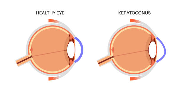
A hyphema is bleeding into the front chamber of the eye frequently caused by trauma. Proper management is essential to avoid complications such as increased eye pressure or corneal staining.
- Presenting signs : Visible blood pooling inside the anterior chamber leads to blurred vision, discomfort, and sensitivity to light .
- Diagnostic approach: Slit-lamp examination assesses bleed size , presence of clot , intraocular pressure measurements monitor secondary glaucoma development .
- Therapeutic strategies: Includes head elevation , protective eyewear , activity restriction , topical medications , sometimes surgical evacuation if bleeding persists .
- Prognosis considerations: Most mild hyphemas resolve over days; severe cases prompt close follow-up due to risk for vision-threatening sequelae .
Immediate consultation after an eye injury causing hyphema enables tailored treatment minimizing long-term complications .
CENTRAL RETINAL ARTERY OCCLUSION

This condition involves sudden blockage of blood flow through the central retinal artery causing acute painless vision loss. It requires urgent evaluation by an eye specialist.
- Main features:Painless loss of vision in one eye often happens without warning due to embolism or thrombosis obstructing arterial flow.
- Diagnostic methods:Dilated fundus exam reveals characteristic pale retina with a cherry red spot at the macula confirming diagnosis.
- Treatment options:Eme rgency interventions include ocular massage, lowering intraocular pressure, hyperbaric oxygen therapy or systemic therapies aimed at restoring circulation where possible.
- Counseling on vascular risk factors helps prevent recurrences beyond acute management milestones.
Rapid identification paired with immediate medical response offers best chance to preserve some visual function following central retinal artery occlusion.
CHEMICAL EYE BURN MANAGEMENT
Chemical burns to the eye require immediate irrigation and care to limit damage. Such injuries are considered ocular emergencies demanding swift action.
- Causative agents: Acids or alkalis can cause superficial or deep tissue injury depending on exposure duration and concentration.
- Initial assessment: Thorough history-taking alongside measurement of pH level guides treatment urgency and approach.
- Treatment protocols: Continuous eye washing followed by medications like antibiotics, steroids, and pain control promote healing.
- Prognosis factors: Timely decontamination improves outcomes although severe burns may result in lasting visual impairment or scarring.
If you suspect a chemical injury to your eyes, immediate rinsing with clean water before professional care substantially reduces long-term harm risks.
RETINAL DETACHMENT REPAIR
A retinal detachment occurs when the retina separates from its underlying supportive tissue. This can rapidly lead to vision loss if not promptly managed.
- Symptoms and causes: Patients may notice sudden flashes of light, floaters, or a shadow over part of their vision caused by trauma or degenerative changes.
- Diagnosis: Comprehensive eye examination with dilated fundoscopy identifies the detached retina location and extent.
- Treatment options: Surgical procedures such as scleral buckling or vitrectomy reattach the retina to restore normal anatomy.
- Recovery process: Post-surgical positioning and follow-up visits ensure proper healing and monitor for complications.
The chances of regaining useful sight increase significantly with timely surgical repair after retinal detachment.
ACUTE ANGLE-CLOSURE GLAUCOMA
Acute angle-closure glaucoma is a sudden increase in eye pressure that can cause rapid vision loss if untreated. It is an ophthalmic emergency requiring prompt attention.
- Symptoms and causes: Severe eye pain, headache, nausea, blurry vision, and halos around lights often occur due to blockage of fluid drainage in the eye.
- Diagnosis: Measurement of intraocular pressure and examination of the anterior chamber angle help confirm this condition.
- Treatment options: Immediate medication to lower pressure, laser therapy, or surgical intervention are used to prevent optic nerve damage.
- Management expectations: Early treatment typically stabilizes pressure and preserves vision; delays may result in permanent impairment.
Recognizing symptoms early and seeking urgent care is critical for successful outcomes in acute angle-closure glaucoma cases.