
Dr Amit M Vora
About Me
Dr Amit M Vora
Cardiologist
Biography
My Clinic
OPD Hours
| Tuesday | Morning | 10:00 AM | - | 02:00 PM |
| Evening | 06:00 PM | - | 09:00 PM | |
| Wednesday | Morning | 10:00 AM | - | 02:00 PM |
| Evening | 06:00 PM | - | 09:00 PM | |
| Thursday | Morning | 10:00 AM | - | 02:00 PM |
| Evening | 06:00 PM | - | 09:00 PM | |
| Friday | Morning | 10:00 AM | - | 02:00 PM |
| Evening | 06:00 PM | - | 09:00 PM | |
| Saturday | Morning | 10:00 AM | - | 02:00 PM |
| Evening | 06:00 PM | - | 09:00 PM | |
| Sunday | Morning | 10:00 AM | - | 02:00 PM |
| Evening | 06:00 PM | - | 09:00 PM | |
| Thursday | Morning | 10:00 AM | - | 02:00 PM |
| Evening | 06:00 PM | - | 09:00 PM |
Treatments
Specialised care and treatments offered
ELECTROPHYSIOLOGICAL STUDY (EPS)

The electrophysiological study (EPS) investigates your heart's electrical system through precise measurements. This diagnostic tool identifies arrhythmias—irregular heartbeats—providing crucial insight into their origins and mechanisms while also guiding treatment choices effectively following careful analysis conducted by trained specialists engaged within varied medical institutions worldwide today!
- Symptoms: Palpitations feeling irregular beats accompanied sometimes by lightheadedness fading awareness signify need arise here indicating possible underlying dysfunction observed via appropriate testing protocols tailored each individual case examined exhaustively without leaving any details uncovered analyzing past medical histories too ultimately clarifying next steps taken accordingly.
Diagnosis: Beginning with physical examinations revealing these alerts considering prior documentation reviewed immediately leads specialists recommending proceeding ahead towards performing EPS setups ensuring maximum detail captured throughout recordings made along pathway observed finally yielding helpful insights toward future recommendations suggested per results obtained afterwards letting things unfold seamlessly maintaining clarity everything efficiently managed ultimately excessively reassuring important matters concerning patients’ health awareness levels remain optimal facilitated ongoing guidance required following conclusion-phase reviews conducted post-checkup sessions too especially thereafter reflecting combined efforts exerted cumulatively altogether working together harmoniously!
Treatment/Management Options:: Treatment plans initiated depend largely upon data analyzed upon conclusion outputs yielded via electrophysiology evaluations prepared thoughtfully professionals dedicated ensuring granular needs met quickly paving pathways suited uniquely according distinctive requirements awaited concluding remarks shared across channels considered vital benefitting stakeholders involved undoubtedly whether medically inclined specialists themselves OR concerned family members expecting clarity coming forth deeply explored emphasizing communication top priority maintained here!
TRANSESOPHAGEAL ECHOCARDIOGRAM (TEE)
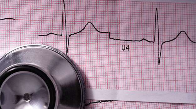
A transesophageal echocardiogram (TEE) provides detailed images of your heart through an ultrasound probe inserted into the esophagus. This method offers advantages over standard echocardiograms in certain clinical situations where clearer images are needed.
- This test may be recommended if patients experience specific symptoms such as unexplained atrial fibrillation or congenital heart defects revealed in earlier assessments.
- Differential diagnosis usually includes thorough discussions regarding symptoms identified during previous exams that prompt further exploration into cardiac structures and function via TEE imagery materials processed by specialized equipment available in most cardiology centers today.
Treatment approaches might differ considerably based on findings from such comprehensive examinations assisting clinicians in timely interventions.
Preparation typically involves fasting before undergoing this procedure which also tends not to require long recovery times afterward enhancing patient comfort significantly throughout their visit as well.
CORONARY ANGIOGRAPHY
Coronary angiography is an imaging technique used to visualize the inside of coronary arteries. It plays a critical role in identifying blockages that could lead to serious cardiovascular events like heart attacks.
- Typical symptoms, such as severe chest pain or unusual shortness of breath during activities, often prompt this investigation after initial assessments indicate risk factors for coronary artery disease (CAD).
- This invasive procedure begins with prior diagnostic evaluations including blood tests and medical history assessments to determine necessity.
- Treatment may subsequently involve angioplasty or stenting if significant blockages are found during the procedure; otherwise, lifestyle modifications might be recommended.
Understanding this process helps reassure patients as they prepare for necessary cardiac evaluations aimed at maintaining optimal heart health.
HOLTER MONITOR
A Holter monitor is a portable device that continuously records your heart's rhythms for 24 to 48 hours. It captures data that helps diagnose irregularities that may not appear during routine exams.
- Patients often seek this test due to symptoms like dizziness, fainting spells, or unexplained palpitations that occur intermittently.
- This diagnostic tool involves wearing electrodes attached to your chest which connect to the monitor throughout its duration.
- If any abnormal rhythms are detected, treatment may vary from lifestyle changes to medication based on findings from the recorded data.
- The monitor allows normal activities during its use, providing real-world insight into how your heart functions under daily conditions.
The Holter monitor is an invaluable resource for tracking transient symptoms and informing subsequent care strategies effectively.
CARDIAC STRESS TEST
A cardiac stress test evaluates how well your heart performs under physical stress. It's crucial in diagnosing various heart conditions and assessing overall cardiovascular health.
- Common symptoms prompting a stress test include unexplained fatigue, palpitations, or chest discomfort during exercise.
- This test is diagnosed primarily through patient history and physical exams that reveal signs of potential heart disease.
- Management may involve medication adjustments or recommendations for lifestyle changes based on your performance during the test.
- The process usually involves walking on a treadmill or pedaling on a stationary bike while monitoring your heart rate and rhythm closely.
- Your healthcare team will guide you throughout; it's essential to understand what to expect before undergoing this evaluation.
A cardiac stress test helps pinpoint issues and tailor appropriate treatment plans for individuals concerned about their heart health.
ECHOCARDIOGRAM
An echocardiogram is a non-invasive imaging test that uses sound waves to create pictures of the heart. It provides valuable information about the size and shape of the heart, as well as how well it’s functioning.
- Symptoms indicating the need for an echocardiogram may include chest pain, shortness of breath, or abnormal heart rhythms.
- The test is typically diagnosed through a physical examination followed by this imaging study when cardiovascular issues are suspected.
- Treatment or management options derived from the results can range from lifestyle changes to surgical interventions based on findings.
- During the procedure, patients can expect a comfortable environment; you’ll lie down while a technician applies gel and moves a transducer over your chest.
- No recovery time is necessary as this is a painless procedure with no known risks involved.
An echocardiogram is essential for understanding heart conditions and guiding treatment decisions effectively.