
Dr Ashok Puri M
About Me
Dr Ashok Puri M
DO
Ophthalmologist
10 Years Experience
Biography
My Clinic
OPD Hours
| Monday | Morning | 10:00 AM | - | 02:00 PM |
| Evening | 06:00 PM | - | 09:00 PM | |
| Tuesday | Morning | 10:00 AM | - | 02:00 PM |
| Evening | 06:00 PM | - | 09:00 PM | |
| Wednesday | Morning | 10:00 AM | - | 02:00 PM |
| Evening | 06:00 PM | - | 09:00 PM | |
| Thursday | Morning | 10:00 AM | - | 02:00 PM |
| Evening | 06:00 PM | - | 09:00 PM | |
| Friday | Morning | 10:00 AM | - | 02:00 PM |
| Evening | 06:00 PM | - | 09:00 PM | |
| Saturday | Morning | 10:00 AM | - | 02:00 PM |
| Evening | 06:00 PM | - | 09:00 PM | |
| Sunday | Morning | 10:00 AM | - | 02:00 PM |
| Evening | 06:00 PM | - | 09:00 PM |
Treatments
Specialised care and treatments offered
OCULAR TRAUMA - BLUNT INJURY
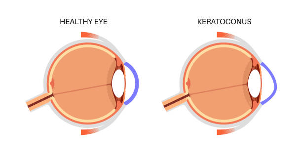
The eye can sustain blunt force trauma leading to a spectrum of injuries needing careful evaluation. Early detection prevents secondary complications effectively.
- User symptoms & risks:: Sudden pain, decreased vision, swelling around the eye often caused by sports injuries or accidents involving impact without penetration.
- Evaluation approach:: Visual acuity testing along with slit lamp exam and imaging (if needed) to rule out globe rupture or vitreous hemorrhage are essential assessments. Treatment options: Management ranges from observation for mild bruising to surgical repair for orbital fractures or intraocular bleeding depending on findings;
This content does not mention that last bullet point properly formatted - will fix below>
CHEMICAL EYE INJURY

The eye’s exposure to harmful chemicals can inflict rapid damage requiring emergency measures. Prompt action reduces potential long-term harm considerably.
- Main symptoms and sources: Painful red eye, watering, reduced vision following contact with acids or alkalis during household or industrial accidents.
- Assessment procedures: Clinical inspection under slit lamp to grade injury severity including corneal epithelial defects and chemical penetration depth assessment.
- Treatment steps: Immediate and copious irrigation with saline or water followed by topical antibiotics, anti-inflammatory agents and frequent monitoring for complications like increased intraocular pressure or infection.
- Counseling on prognosis: Mild injuries usually heal well but severe exposures risk scarring requiring reconstructive surgery such as amniotic membrane transplantation later on.
RETINAL DETACHMENT
The separation of the retina from its underlying tissue demands swift diagnosis to prevent permanent vision loss. Understanding warning signs enables timely medical attention.
- Symptoms and causes: Sudden appearance of floaters, flashes of light, or a shadow/curtain in peripheral vision; frequently linked with trauma or degenerative retinal changes.
- Diagnosis methods: A comprehensive dilated fundus examination using indirect ophthalmoscopy reveals retinal tears or detachment extent.
- Treatment modalities: Surgical intervention such as pneumatic retinopexy, scleral buckle, or vitrectomy is typically required depending on detachment severity and location.
- Treatment outcomes: Early surgery greatly enhances the likelihood of reattachment and visual recovery; delayed repair risks permanent blindness in affected areas.
This condition is an ocular emergency where immediate specialist evaluation determines visual prognosis greatly.
CORNEAL ULCER

An infection affecting the cornea can cause serious visual impairment if untreated. Protective measures and timely treatment reduce complications substantially.
- Symptoms and causes: Redness, severe eye pain, tearing, discharge, blurred vision; commonly results from bacterial, fungal or viral infections often after trauma or contact lens use.
- Diagnostic approach: Slit-lamp examination and microbial cultures of corneal scrapings help identify causative agents precisely.
- Treatment strategies: Intensive topical antimicrobial drops tailored to organism sensitivity; sometimes oral therapy or hospitalization is required for severe cases.
- Follow-up expectations: Healing may take weeks with gradual symptom resolution; scarring risk necessitates ongoing evaluation.
Aggressive management at first presentation improves prognosis significantly in these potentially sight-threatening infections.
ACUTE ANGLE-CLOSURE GLAUCOMA
Sudden elevation of eye pressure due to blocked drainage can cause a medical emergency requiring prompt attention. Recognizing the signs early is crucial to prevent lasting damage.
- Symptoms and causes: Intense eye pain, headache, nausea, blurred vision, halos around lights; caused by rapid closure of the anterior chamber angle.
- Diagnosis: Measurement of intraocular pressure using tonometry and gonioscopy to inspect the drainage angle.
- Treatment options: Immediate administration of pressure-lowering medications may be followed by laser or surgical iridotomy to restore fluid flow.
- Management: Close monitoring after treatment is vital as recurrence can occur; patients should avoid medications that can worsen angle closure.
Early recognition and intervention drastically improve outcomes and preserve vision. Understanding symptoms helps patients seek urgent care promptly.