
Dr Bhawna Sharma
About Me
Dr Bhawna Sharma
MD
Neurologist
10 Years Experience
Biography
My Clinic
OPD Hours
| Monday | Morning | 10:00 AM | - | 02:00 PM |
| Evening | 06:00 PM | - | 09:00 PM | |
| Tuesday | Morning | 10:00 AM | - | 02:00 PM |
| Evening | 06:00 PM | - | 09:00 PM | |
| Wednesday | Morning | 10:00 AM | - | 02:00 PM |
| Evening | 06:00 PM | - | 09:00 PM | |
| Thursday | Morning | 10:00 AM | - | 02:00 PM |
| Evening | 06:00 PM | - | 09:00 PM | |
| Friday | Morning | 10:00 AM | - | 02:00 PM |
| Evening | 06:00 PM | - | 09:00 PM | |
| Saturday | Morning | 10:00 AM | - | 02:00 PM |
| Evening | 06:00 PM | - | 09:00 PM | |
| Sunday | Morning | 10:00 AM | - | 02:00 PM |
| Evening | 06:00 PM | - | 09:00 PM |
Treatments
Specialised care and treatments offered
CT SCAN OF THE BRAIN

A CT scan, or computed tomography scan of the brain provides detailed cross-sectional images useful for diagnosing various conditions such as strokes, tumors, head injuries, and more. It utilizes X-ray technology combined with computer processing for clarity and precision in imaging results.
- Symptoms Leading To CT Scans: Sudden severe headaches, confusion, vision problems are common triggers indicating this examination might be needed.
- The Scanning Process: Patients lie down within a tube-like machine while several X-ray beams rotate around them capturing slices of images from different angles.
- What Can Be Diagnosed? : Findings can clarify whether there’s bleeding within cranial spaces or identify mass lesions effectively guiding urgent care decisions when necessary.
- Safety Aspects: Although exposure occurs during scanning due to radiation use—benefits vastly outweigh risks when performed prudently under medical guidance!
Performing CT scans helps clinicians ascertain immediate factors affecting patient health when neurologic concerns arise rapidly!
CEREBRAL ANGIOGRAPHY
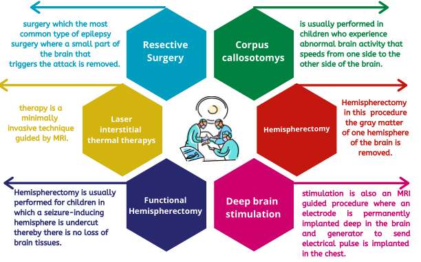
Cerebral angiography is an advanced imaging technique used to visualize blood vessels in and around the brain. This invasive but highly informative test helps diagnose vascular diseases like aneurysms or arteriovenous malformations (AVMs).
- Clinical Indications: It’s commonly recommended for patients experiencing unexplained headaches or stroke symptoms where vascular anomalies are suspected.
- Diagnostic Method: A catheter is threaded through blood vessels leading to cerebral arteries; contrast dye highlights areas on X-ray images.
- Treatment Implications: Results assist in determining whether surgical intervention or other treatments are necessary for vascular lesions detected.
- Patient Experience: Though it involves some risks due to its invasive nature, monitoring makes it reasonably safe under professional care.
This diagnostic approach plays an essential role in managing complex cerebrovascular conditions effectively.
NERVE CONDUCTION STUDY
A Nerve Conduction Study (NCS) evaluates how fast signals travel through peripheral nerves. This test is critical for diagnosing nerve damage or disorders affecting nerve function such as carpal tunnel syndrome or neuropathies.
- Main Symptoms: Weakness, pain, tingling sensations in extremities are potential indicators requiring this assessment.
- The Testing Process: Electrodes are placed on the skin over nerves; small electrical impulses stimulate nerves while their responses are recorded.
- Treatment Planning:If abnormalities are found, treatment options may include physical therapy or medications for symptom relief.
- Duration & Comfort: Generally takes about 30-60 minutes; most individuals report only minor discomfort from electrode placement.
By assessing nerve function accurately, this study aids in identifying underlying causes of sensory and motor symptoms.
LUMBAR PUNCTURE
The lumbar puncture, often referred to as a spinal tap, is a procedure used to collect cerebrospinal fluid (CSF) for analysis. This fluid surrounds the brain and spinal cord, providing vital information about various neurological conditions such as infections or multiple sclerosis.
- Symptoms Indicating Need: Patients with symptoms like severe headaches or signs of infection may be advised to undergo this procedure.
- The Procedure: A thin needle is inserted into the lower back area between two vertebrae to obtain CSF samples safely.
- Differential Diagnosis: Analyzing CSF can help identify infections, bleeding around the brain, or other central nervous system disorders.
- Anesthesia Used: Local anesthesia minimizes discomfort during the procedure; most patients report only mild pressure sensations.
This vital diagnostic tool enhances understanding of complex neurological conditions and informs subsequent management strategies.
MAGNETIC RESONANCE IMAGING (MRI)
A magnetic resonance imaging (MRI) scan is a powerful imaging technique used to visualize the structures of the brain and spinal cord. This non-invasive method uses magnetic fields and radio waves to create detailed images that can reveal abnormalities such as tumors, trauma, or degenerative diseases.
- Common Indications: Symptoms prompting an MRI may include persistent headaches, dizziness, or neurological deficits.
- How it Works: Patients lie inside a large cylindrical machine while it captures high-resolution images of brain tissues.
- Treatment Insights: Findings can guide treatment decisions ranging from medication management to surgical interventions.
- Painless Experience: The process is typically quiet and does not involve radiation; however, some patients may experience claustrophobia.
This imaging modality plays a crucial role in diagnosing diverse neurological conditions effectively.
ELECTROENCEPHALOGRAM (EEG)
An electroencephalogram (EEG) is a diagnostic test that measures electrical activity in the brain. It is primarily used to evaluate conditions such as seizures, epilepsy, and other neurological disorders. This non-invasive procedure involves placing electrodes on the scalp to record brain waves.
- Symptoms/Conditions: Patients may experience seizures, periods of confusion, or unexplained loss of consciousness.
- Diagnostic Process: An EEG is performed in a clinical setting where electrodes are attached; the results help determine the presence and type of abnormal brain activity.
- Treatment Options: Depending on findings, treatment may include medications to control seizures or further neurological assessments.
- What to Expect: The procedure is painless and typically lasts 30-60 minutes, with results available shortly after completion.
This essential diagnostic tool assists healthcare providers in formulating an appropriate treatment plan for patients suffering from various neurological issues.