
Dr Bhupender Sin
About Me
Dr Bhupender Sin
DO
Ophthalmologist
10 Years Experience
Biography
My Clinic
OPD Hours
| Monday | Morning | 10:00 AM | - | 02:00 PM |
| Evening | 06:00 PM | - | 09:00 PM | |
| Tuesday | Morning | 10:00 AM | - | 02:00 PM |
| Evening | 06:00 PM | - | 09:00 PM | |
| Wednesday | Morning | 10:00 AM | - | 02:00 PM |
| Evening | 06:00 PM | - | 09:00 PM | |
| Thursday | Morning | 10:00 AM | - | 02:00 PM |
| Evening | 06:00 PM | - | 09:00 PM | |
| Friday | Morning | 10:00 AM | - | 02:00 PM |
| Evening | 06:00 PM | - | 09:00 PM | |
| Saturday | Morning | 10:00 AM | - | 02:00 PM |
| Evening | 06:00 PM | - | 09:00 PM | |
| Sunday | Morning | 10:00 AM | - | 02:00 PM |
| Evening | 06:00 PM | - | 09:00 PM |
Treatments
Specialised care and treatments offered
CORNEAL TOPOGRAPHY
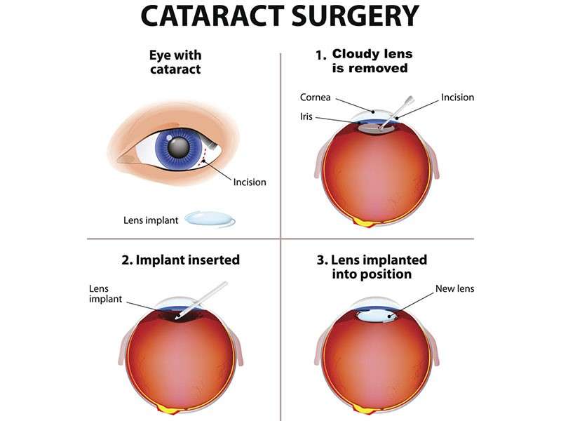
A corneal topography test produces detailed maps revealing the curvature shape and surface features of the cornea—the transparent front window of the eye. It supports diagnosis and management planning for multiple corneal disorders and refractive surgery assessments.
- CLINICAL IMPORTANCE : Crucial in detecting keratoconus (progressive thinning & bulging), irregular astigmatism or planning LASIK/PRK procedures safely
- TECHNOLOGY : Uses computerized imaging systems capturing thousands of data points reflecting surface irregularities accurately within minutes
- PATIENT EXPERIENCE : Non-invasive; requires steady fixation during rapid image acquisition without direct contact
- APPLICATIONS : Helps monitor disease progress over time & guides fitting specialty contact lenses improving visual outcomes significantly
This advanced diagnostic approach provides vital information influencing precision care decisions tailored uniquely based on individual corneal architecture details.
TONOMETRY
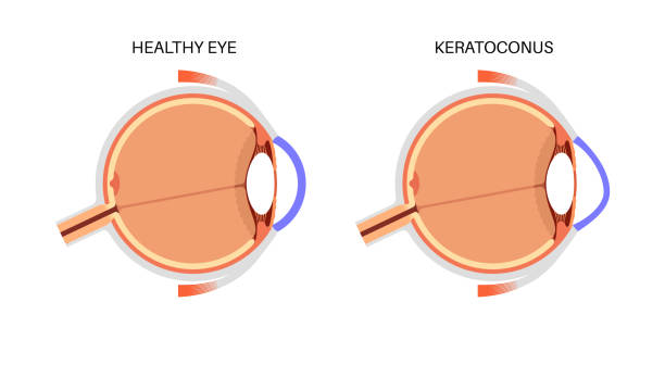
Tonometry measures intraocular pressure (IOP), which is critical in assessing risk or presence of glaucoma—a condition characterized by damage to the optic nerve often associated with elevated pressure inside the eye.
- TYPES OF TONOMETRY: Includes Goldmann applanation tonometry (gold standard), non-contact air-puff tonometry, and others tailored to patients' needs and clinical settings.
- SIGNIFICANCE OF IOP MEASUREMENT:
FUNDUS PHOTOGRAPHY
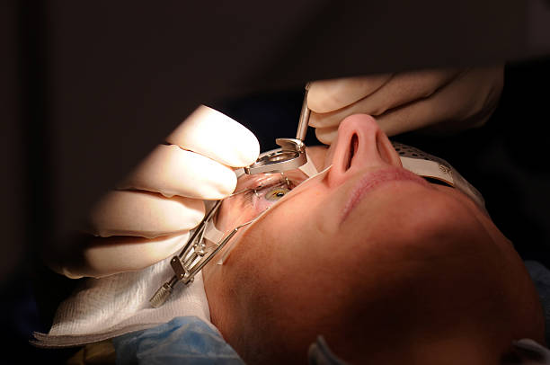
Fundus photography involves capturing detailed images of the retina, optic disc, macula, and posterior pole using specialized cameras. These images assist clinicians in documenting ocular health and pathology progression over time.
- Main Uses: Monitoring diabetic retinopathy, macular degeneration, hypertensive changes, and optic neuropathies effectively.
- The Process: Non-invasive photo session typically with pupil dilation to enhance image clarity and extent coverage.
- Efficacy: Enables visualization of subtle retinal lesions invisible during ordinary examination techniques alone.
- Treatment Role: Assists ophthalmologists with objective documentation facilitating comparisons across visits for personalized care adjustments as needed.
VISUAL FIELD TESTING
The visual field test evaluates peripheral vision to detect blind spots or defects that could indicate underlying ocular or neurological issues. It's pivotal in glaucoma care and other optic nerve pathologies.
- Sensation Assessment: Measures the scope of vision beyond central focus areas by presenting light stimuli at different points.
- Disease Detection: Early peripheral deficits help diagnose glaucoma before noticeable vision loss occurs.
- User Experience: Usually requires patient attention to respond when lights are seen during testing sessions lasting 10-20 minutes.
- Diversified Methods: Includes automated techniques like Humphrey perimetry offering reliable reproducibility of results.
This screening tool is crucial for comprehensive eye exams aimed at preserving long-term visual function through timely intervention strategies.
OPTICAL COHERENCE TOMOGRAPHY (OCT)
Optical Coherence Tomography (OCT) is an advanced imaging technique that captures cross-sectional images of the retina with high resolution. It plays a key role in diagnosing various retinal conditions.
- Function: Creates detailed maps of retinal layers to identify structural abnormalities.
- Indications: Commonly used for macular degeneration, diabetic retinopathy, and glaucoma monitoring.
- Process: Non-invasive scan takes just a few minutes without discomfort or eye contact.
- Advantages: Provides quantitative data to track disease progression over time objectively.
- Treatment Guidance: Results assist ophthalmologists in tailoring appropriate therapy plans effectively.
This technology greatly enhances early diagnosis and monitoring accuracy for various sight-threatening diseases.
SLIT LAMP EXAMINATION
A slit lamp examination is an essential diagnostic procedure that allows detailed inspection of the eyes' anterior and posterior segments. It utilizes a specialized microscope providing a magnified, three-dimensional view.
- Purpose: Helps detect abnormalities in the cornea, iris, lens, and retina.
- Procedure: The patient rests their chin on a support while the clinician examines the eye under focused light.
- Diagnoses: Useful for identifying cataracts, corneal injuries, infections, and glaucoma signs.
- Preparation: Usually no special preparation needed; sometimes pupil dilation may be necessary.
- Outcome: This painless test aids in early detection of eye diseases for timely management.
This examination forms a cornerstone in routine ophthalmic evaluations and helps guide further treatments or investigations if required.