
Dr Deepak Shetty
About Me
Dr Deepak Shetty
Cardiologist
Biography
My Clinic
OPD Hours
| Tuesday | Morning | 10:00 AM | - | 02:00 PM |
| Evening | 06:00 PM | - | 09:00 PM | |
| Wednesday | Morning | 10:00 AM | - | 02:00 PM |
| Evening | 06:00 PM | - | 09:00 PM | |
| Thursday | Morning | 10:00 AM | - | 02:00 PM |
| Evening | 06:00 PM | - | 09:00 PM | |
| Friday | Morning | 10:00 AM | - | 02:00 PM |
| Evening | 06:00 PM | - | 09:00 PM | |
| Saturday | Morning | 10:00 AM | - | 02:00 PM |
| Evening | 06:00 PM | - | 09:00 PM | |
| Sunday | Morning | 10:00 AM | - | 02:00 PM |
| Evening | 06:00 PM | - | 09:00 PM | |
| Thursday | Morning | 10:00 AM | - | 02:00 PM |
| Evening | 06:00 PM | - | 09:00 PM |
Treatments
Specialised care and treatments offered
STRESS TESTING

A stress test evaluates how well your heart performs under physical exertion. It measures responses when subjected to activities akin to everyday tasks; making this crucial for diagnosing potential cardiovascular issues effectively.
- Commonly monitored signs include changes in blood pressure , electrocardiogram results , and overall tolerance levels throughout testing stages .
- Symptoms warranting may involve chest discomfort , abnormal heartbeat sensations , unexplained fatigue during physical activities over time .
- Stress tests can come in various forms: treadmill tests (exercise-based) & pharmacologic methods using medications induce similar effects safely without requiring actual exercise capabilities .
- Test lengths vary depending upon protocol implemented but last roughly around 15 – 20 minutes typically tailoring towards patient circumstances encountered providing equally valuable insights nonetheless .
Understanding performance dynamics aids physicians promoting proactive preventative measures linking treatments/care responsive towards overall long-term wellness objectives tracking progress continually addressing concerns raised iteratively considering evolving health status beyond initial assessments preemptively fortifying resilience against future complications possibly arising altogether!
TRANSESOPHAGEAL ECHOCARDIOGRAM (TEE)
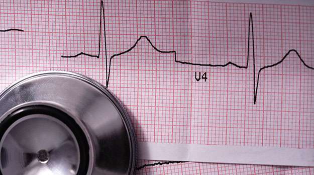
A transesophageal echocardiogram (TEE) is an advanced imaging technique used to obtain clearer images of the heart's structures compared to traditional echocardiograms. TEE involves inserting a flexible probe down the esophagus, providing direct access nearby without interference from lung tissue or ribs.
- This procedure is especially useful for diagnosing blood clots , valvular diseases , and congenital anomalies in complex cases where standard imaging falls short.
- Patients typically require fasting before this test because sedation will be administered.
- The test generally takes between 30 minutes to an hour , allowing clinicians precise evaluations while minimizing discomfort.
- Post-procedure recovery includes monitoring until sedation wears off but often allows patients home shortly after with minimal downtime involved .
TEE serves as an important tool that enhances diagnostic accuracy while assisting in forming effective management plans tailored toward individual therapeutic needs .
ELECTROPHYSIOLOGY STUDY
An electrophysiology study (EPS) is performed to evaluate the electrical system of the heart. This procedure helps diagnose arrhythmias by mapping electrical impulses and identifying areas contributing to abnormal rhythms or conduction delays.
- Patiens may experience symptoms such as rapid heartbeat, dizziness, or fainting spells leading up to this diagnostic test.
- The procedure entails inserting catheters through blood vessels into the heart under sedation; it usually lasts several hours depending on findings.
- A thorough investigation allows doctors to determine whether interventions such as ablation or device implantation are necessary for rhythm correction.
- This minimally invasive approach provides critical insights while also offering therapeutic options if needed during the same session.
An EPS can significantly improve outcomes for patients suffering from persistent arrhythmias by guiding appropriate treatment strategies based on specific findings from their unique cases.
CARDIAC MONITORING
Cardiac monitoring allows physicians to observe a patient's heart activity over an extended period. This service can be critical for diagnosing arrhythmias or other electrical disturbances within the heart that might not be evident during a standard examination.
- Common reasons for cardiac monitoring include episodes of palpitations, dizziness, or syncope (fainting).
- A variety of techniques exist, such as Holter monitors worn for 24-48 hours or event recorders that capture data during symptomatic episodes.
- The evaluation process helps identify abnormal rhythms that may lead to serious conditions like atrial fibrillation or ventricular tachycardia.
- No invasive procedures are involved—patients simply wear a device while going about their daily activities for real-time analysis.
- The results enable tailored management approaches ranging from lifestyle modifications to potential interventions if needed.
This advanced monitoring provides invaluable information regarding patient-specific cardiac health without significant disruption to daily life.
ECHOCARDIOGRAM
An echocardiogram is a non-invasive imaging test that employs ultrasound waves to create detailed images of the heart's structure and function. This essential tool helps in diagnosing various heart conditions, providing insights into how the heart is working.
- This test can reveal issues such as valve diseases, congenital heart defects, and signs of previous heart attacks.
- Echocardiograms are often ordered following symptoms like unexplained fatigue or shortness of breath.
- The procedure involves placing small sensors on the chest area that capture sound waves; it usually lasts about 30-60 minutes.
- No special preparation is required, making it a straightforward process for patients.
- The results help guide treatment plans, which may include medication adjustments, lifestyle changes, or further testing if necessary.
This comprehensive assessment enables healthcare providers to monitor heart health effectively over time.
CORONARY ANGIOPLASTY
Coronary angioplasty is a minimally invasive procedure designed to open up narrowed or blocked coronary arteries. This treatment is primarily aimed at improving blood flow to the heart muscle and alleviating symptoms of coronary artery disease.
- Symptoms may include chest pain (angina), shortness of breath, and fatigue during physical activities.
- Diagnosis typically involves angiography, an imaging technique that utilizes contrast dye and X-rays to visualize blood flow in the coronary arteries.
- Treatment can involve placing a stent after balloon inflation to keep the artery open, thus preventing future blockages.
- The procedure usually takes about one hour and is performed in a catheterization lab under local anesthesia.
- Post-procedure care includes monitoring for any complications and prescribing medication to prevent clots.
Coronary angioplasty is often effective in relieving symptoms and improving quality of life for patients with significant coronary artery obstruction.