
Dr Dilpreet Singh
About Me
Dr Dilpreet Singh
MS
Ophthalmologist
10 Years Experience
Biography
My Clinic
OPD Hours
| Monday | Morning | 10:00 AM | - | 02:00 PM |
| Evening | 06:00 PM | - | 09:00 PM | |
| Tuesday | Morning | 10:00 AM | - | 02:00 PM |
| Evening | 06:00 PM | - | 09:00 PM | |
| Wednesday | Morning | 10:00 AM | - | 02:00 PM |
| Evening | 06:00 PM | - | 09:00 PM | |
| Thursday | Morning | 10:00 AM | - | 02:00 PM |
| Evening | 06:00 PM | - | 09:00 PM | |
| Friday | Morning | 10:00 AM | - | 02:00 PM |
| Evening | 06:00 PM | - | 09:00 PM | |
| Saturday | Morning | 10:00 AM | - | 02:00 PM |
| Evening | 06:00 PM | - | 09:00 PM | |
| Sunday | Morning | 10:00 AM | - | 02:00 PM |
| Evening | 06:00 PM | - | 09:00 PM |
Treatments
Specialised care and treatments offered
CORNEAL TOPOGRAPHY
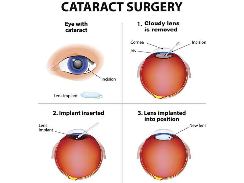
Corneal topography captures detailed maps of the cornea's surface curvature. By understanding these patterns provides insights into various conditions such as keratoconus or astigmatism. It plays a critical role in ensuring accurate fitting for contact lenses as well as planning refractive surgeries like LASIK
.- Sensitivity & Symptoms: Common indicators warranting this evaluation might include distorted vision or frequent prescription changes
- Tests combine computer analysis alongside specialized devices tracking each curve both pre- & post-surgery ensuring maximum precision throughout procedures Post-Testing: Normal follow-ups ascertain optimal recovery durations tailored per patient improving outcomes overall .
Staying one step ahead concerning wearers preferences manages future alterations promoting healthier routines!
OCULAR ULTRASOUND
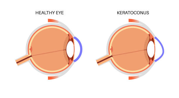
An ocular ultrasound utilizes sound waves to produce images of the structures inside the eye. This technique is particularly useful when other imaging methods are inadequate or when assessing complex issues like tumors or retinal detachments hidden from direct view during examination.
- This painless procedure generally takes only minutes and requires no special preparation by patients beforehand
- Pediatric assessments often benefit from ultrasound due to children’s challenges with standard imaging techniques like dilated fundus exams
This diagnostic tool serves as an integral component in comprehensive ocular evaluations—especially valuable in emergencies!
VISUAL FIELD TESTING
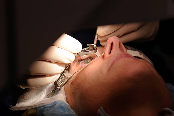
Visual field testing measures your entire scope of vision, including peripheral sight. It helps diagnose conditions such as glaucoma or neurological disorders affecting vision. Understanding your visual field is vital in maintaining overall ocular health.
- Poor peripheral vision may occur without noticeable symptoms until significant damage has occurred, making testing crucial for early detection.
- The test usually involves focusing on a fixed point while lights flash in different areas of your peripheral view; responses help create a visual map of your sight capability.
- Your ophthalmologist will analyze the results to identify any abnormalities related to potential diseases affecting your eyesight.
This simple yet effective test can lead to timely interventions that safeguard against progressive vision loss over time. Maintaining regular testing intervals is essential for those at higher risk
RETINAL IMAGING
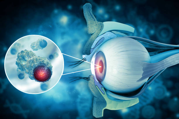
Retinal imaging is a non-invasive diagnostic tool used to capture detailed images of the retina's interior. This technology facilitates early detection of various eye diseases, including diabetic retinopathy and macular degeneration.
- This procedure does not require dilation; it provides high-resolution images quickly and comfortably.
- Common indications for retinal imaging include blurred vision or changes in retinal appearance noted during routine exams.
- The results help ophthalmologists make informed decisions regarding treatment options or referrals as necessary.
This innovative approach enhances monitoring strategies for patients with chronic conditions affecting their eyesight. Regular imaging can lead to proactive management of potential problems before they escalate.
GLAUCOMA ASSESSMENT
Glaucoma is a group of eye conditions that cause damage to the optic nerve, often linked to increased pressure in the eye. Early detection is crucial for preventing vision loss. Regular glaucoma assessments are essential for at-risk individuals.
- The most common symptoms include peripheral vision loss, which can be subtle initially.
- A thorough assessment includes measuring intraocular pressure, visual field tests, and inspecting the optic nerve for signs of damage.
- Management options may involve medications, laser treatments, or surgical procedures aimed at reducing intraocular pressure.
- Follow-up visits are vital to monitor disease progression and adjust treatment plans accordingly.
Regular check-ups with an ophthalmologist can significantly contribute to preserving vision for those with glaucoma risk factors.
CATARACT SURGERY
Cataracts are a common condition where the lens of the eye becomes cloudy, leading to blurred vision. This can significantly impact daily activities and quality of life. Cataract surgery is a safe and effective procedure performed to restore clear vision.
- Symptoms include difficulty seeing at night, sensitivity to light, and faded colors.
- Cataracts are diagnosed through a comprehensive eye examination that includes visual acuity tests and dilating the pupils.
- Treatment typically involves surgery to remove the cloudy lens and replace it with an artificial intraocular lens.
- Post-operative care may include using prescribed eye drops and attending follow-up appointments to monitor healing.
- Most patients experience improved vision within days following the procedure.
It’s important to consult with your ophthalmologist for personalized advice and information on cataract management options.