
Dr Gargi Merchant
About Me
Dr Gargi Merchant
Ophthalmologist
Biography
My Clinic
OPD Hours
| Tuesday | Morning | 10:00 AM | - | 02:00 PM |
| Evening | 06:00 PM | - | 09:00 PM | |
| Wednesday | Morning | 10:00 AM | - | 02:00 PM |
| Evening | 06:00 PM | - | 09:00 PM | |
| Thursday | Morning | 10:00 AM | - | 02:00 PM |
| Evening | 06:00 PM | - | 09:00 PM | |
| Friday | Morning | 10:00 AM | - | 02:00 PM |
| Evening | 06:00 PM | - | 09:00 PM | |
| Saturday | Morning | 10:00 AM | - | 02:00 PM |
| Evening | 06:00 PM | - | 09:00 PM | |
| Sunday | Morning | 10:00 AM | - | 02:00 PM |
| Evening | 06:00 PM | - | 09:00 PM | |
| Thursday | Morning | 10:00 AM | - | 02:00 PM |
| Evening | 06:00 PM | - | 09:00 PM |
Treatments
Specialised care and treatments offered
MACULAR DEGENERATION MANAGEMENT
Age-related macular degeneration (AMD) impacts central vision due to deterioration in the macula part of the retina. It's vital for activities requiring fine detail like reading or driving.
- Symptoms often manifest as straight lines appearing wavy or dark spots in one’s central field of view.
- Diagnosis includes detailed retinal examinations using optical coherence tomography (OCT) which helps visualize thin layers within your retina.
- Treatment strategies may involve anti-VEGF injections directly into the eye paired with lifestyle modifications aiming towards improved nutrition essential for retinal health.
- Patients should anticipate regular assessments after initiating treatment alongside guidance on adapting their everyday routines focusing on clarity preservation throughout this journey.
Proactive management makes an immense difference in slowing progression while enhancing quality living experiences significantly despite challenges faced by AMD.
KERATOCONUS DIAGNOSIS
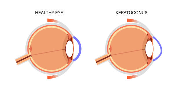
Keratoconus is a progressive condition where the cornea thins out and begins to bulge into a cone-like shape. This irregular curvature distorts vision significantly over time if left untreated.
- The initial signs may include blurriness or difficulty seeing at night due to changing corneal shapes affecting light entry into the eye.
- A specialized corneal topography test measures the curvature of your cornea accurately during diagnosis. This helps visualize any steepening indicative of keratoconus.
- Treatment options range from contact lenses specifically designed for irregular corneas, cross-linking therapy which strengthens corneal tissue, or in advanced cases, surgical intervention such as corneal transplants may be needed.
- Ongoing monitoring ensures any progression of keratoconus is managed effectively with appropriate visual aids or treatments as necessary over time.
Early diagnosis can help slow down its progression greatly while maintaining better visual acuity
RETINAL DETACHMENT ASSESSMENT
A retinal detachment is a serious medical condition that necessitates immediate attention. It occurs when the retina pulls away from its normal position at the back of the eye.
- Common symptoms include sudden flashes of light, floaters, or a shadow over part of your field of vision.
- A comprehensive assessment will involve dilated fundus examination along with imaging techniques like ultrasound if necessary.
- Treatment commonly requires surgical intervention such as scleral buckle or vitrectomy to reattach the retina securely.
- Post-operative care is critical; patients should expect regular follow-ups and adherence to recovery protocols to ensure optimal healing outcomes.
Recognizing symptoms promptly and seeking immediate care can help preserve sight effectively after detachment occurs.
DIABETIC RETINOPATHY EVALUATION
Diabetic retinopathy is a serious complication arising from diabetes that affects the blood vessels in the retina. Regular evaluation is key for early detection and treatment.
- Symptoms may not be noticeable at first but can later include blurred vision, floaters, or even sudden vision loss.
- A thorough eye exam will involve dilating the pupils and taking images of the retina to assess any abnormalities in blood vessels.
- Treatment options vary from laser surgery to injections into the eye depending on the severity of the condition.
- Persistent follow-up appointments are essential for ongoing management and to prevent further complications from diabetes affecting eyesight.
With timely intervention, significant improvements in visual outcomes can be achieved for those diagnosed with this condition.
GLAUCOMA SCREENING
Regular screening for glaucoma is crucial in preventing permanent vision loss associated with this group of eye diseases. Elevated intraocular pressure can damage the optic nerve over time.
- The primary symptoms are often subtle but can include tunnel vision or blind spots as the disease progresses.
- An ophthalmologist will perform tonometry to measure eye pressure as well as other tests such as perimetry to assess peripheral vision.
- If diagnosed, management options may include prescribed medications, laser therapy, or surgery to lower intraocular pressure.
- Patients can expect ongoing monitoring and potential lifestyle adjustments to help manage their condition effectively.
Timely detection plays a vital role in preserving eyesight and maintaining overall eye health.
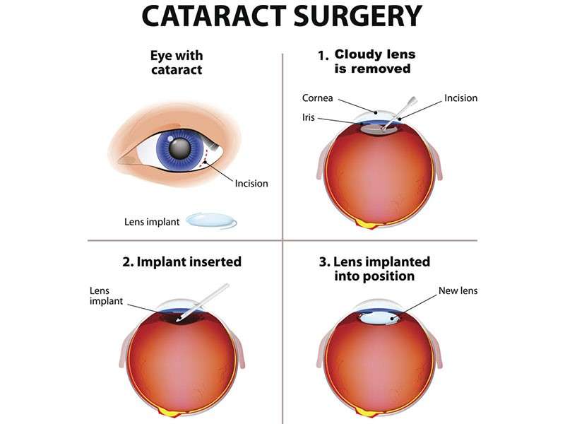
CATARACT SURGERY
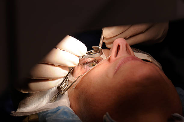
Cataract surgery is a common procedure performed to restore vision for individuals suffering from cataracts. These cloudy formations on the lens of the eye can lead to significant visual impairment.
- Symptoms include blurry vision, difficulty with night vision, and frequent changes in prescription glasses.
- Diagnosis typically involves a comprehensive eye examination, including visual acuity tests and slit-lamp evaluations.
- Treatment consists of surgically removing the cloudy lens and replacing it with an artificial intraocular lens (IOL).
- Post-operative recovery generally involves using prescribed eye drops and attending follow-up appointments to monitor healing.
This procedure has a high success rate, allowing patients to regain clear vision and improve their quality of life.