
Dr Harshut Tak
About Me
Dr Harshut Tak
DO
Ophthalmologist
10 Years Experience
Biography
My Clinic
OPD Hours
| Monday | Morning | 10:00 AM | - | 02:00 PM |
| Evening | 06:00 PM | - | 09:00 PM | |
| Tuesday | Morning | 10:00 AM | - | 02:00 PM |
| Evening | 06:00 PM | - | 09:00 PM | |
| Wednesday | Morning | 10:00 AM | - | 02:00 PM |
| Evening | 06:00 PM | - | 09:00 PM | |
| Thursday | Morning | 10:00 AM | - | 02:00 PM |
| Evening | 06:00 PM | - | 09:00 PM | |
| Friday | Morning | 10:00 AM | - | 02:00 PM |
| Evening | 06:00 PM | - | 09:00 PM | |
| Saturday | Morning | 10:00 AM | - | 02:00 PM |
| Evening | 06:00 PM | - | 09:00 PM | |
| Sunday | Morning | 10:00 AM | - | 02:00 PM |
| Evening | 06:00 PM | - | 09:00 PM |
Treatments
Specialised care and treatments offered
TONOMETRY
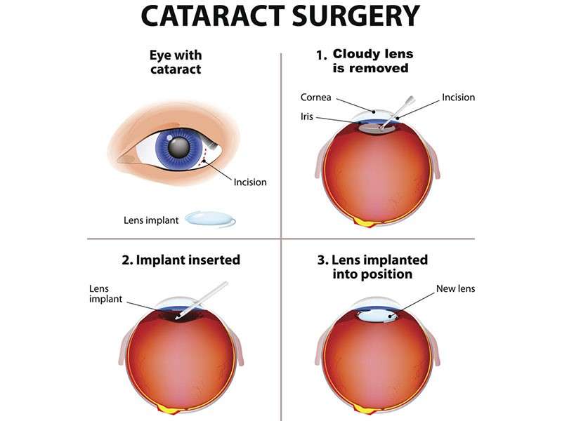
Tonometry measures intraocular pressure (IOP), essential in screening for glaucoma—a leading cause of irreversible blindness globally. Elevated IOP can damage optic nerves if untreated, hence regular measurement is vital especially in at-risk individuals.
- Symptoms/causes : Routine tonometry is suggested particularly for those with family history or symptoms like blurred vision and eye pain .
- Diagnosis : Various techniques exist including air-puff (non-contact) tonometry which gently puffs air onto the eye surface , providing quick readings .
- Treatment/management options : High IOP readings require further glaucoma evaluations followed by medical , laser , or surgical therapies aimed at lowering pressure .
- What to expect : Most methods are painless ; some require numbing drops ; overall , procedure lasts just moments .
Monitoring intraocular pressure regularly through tonometry helps prevent progressive damage preserving eyesight effectively .
CORNEAL TOPOGRAPHY
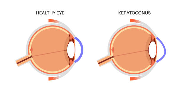
Corneal topography produces detailed maps illustrating the curvature and shape of the cornea's surface. This diagnostic tool is critical in detecting irregularities affecting visual clarity such as keratoconus or fitting contact lenses precisely.
- Symptoms/causes: Indicated when patients experience distorted vision or before refractive surgeries like LASIK to assess suitability.
- Diagnosis: A camera captures reflections off the cornea creating color-coded maps representing elevations and depressions accurately.
- Treatment/management options: Data assists in customizing treatment plans including surgical approaches or specialty lens fittings tailored individually.
- What to expect: The test is quick with no direct contact; it is painless though requires steady head positioning during image acquisition.
This precise mapping enhances outcomes by allowing personalized assessment essential for optimal visual correction strategies.
FUNDUS PHOTOGRAPHY
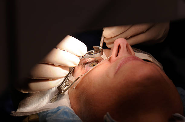
Fundus photography captures detailed images of the interior surface of the eye including the retina, optic disc, and blood vessels. It is invaluable for diagnosing and tracking various retinal conditions over time.
- Symptoms/causes: Often performed during evaluations for diabetes-related eye changes or hypertensive retinopathy screening.
- Diagnosis: Using specialized cameras, photographs are taken after pupil dilation to document current retina health status.
- Treatment/management options: Serial images allow monitoring disease progression guiding timely therapeutic measures like laser treatments if needed.
- What to expect: The process involves minimal discomfort; pupils are dilated using drops beforehand which may cause temporary light sensitivity.
This non-invasive method supports effective long-term eye care through visual documentation of ocular health changes.
VISUAL FIELD TESTING
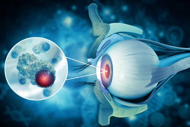
Visual field testing evaluates peripheral vision to detect blind spots or vision loss often unnoticed by patients. It plays a crucial role in diagnosing glaucoma and neurological disorders affecting vision pathways.
- Symptoms/causes: Patients may be referred if they notice tunnel vision or have risk factors for glaucoma or brain lesions.
- Diagnosis: The test involves responding to visual stimuli presented on a screen while focusing on a central point.
- Treatment/management options: Abnormal findings prompt further evaluation and interventions like pressure-lowering medications for glaucoma.
- What to expect: The procedure typically takes 20-30 minutes per eye and requires patient cooperation but is completely painless.
This examination helps preserve vision by assessing functional aspects beyond what can be seen on imaging alone.
OPTICAL COHERENCE TOMOGRAPHY (OCT)
Optical Coherence Tomography (OCT) is an advanced imaging technique that provides cross-sectional images of the retina. It helps assess retinal thickness and identify abnormalities affecting vision.
- Symptoms/causes: Recommended for patients experiencing visual distortions or diagnosed with conditions like macular degeneration or diabetic retinopathy.
- Diagnosis: OCT uses light waves to capture detailed images of retinal layers without any discomfort.
- Treatment/management options: Results assist in monitoring disease progression and tailoring treatments such as injections or laser therapy.
- What to expect: The scan is quick (around 10 minutes), non-invasive, and requires no special preparation.
By providing detailed visualization beneath the retinal surface, OCT enhances early detection and management of sight-threatening diseases.
SLIT LAMP EXAMINATION
A slit lamp examination is a fundamental diagnostic procedure used to evaluate the structures of the eye under high magnification. It allows detailed visualization of both the anterior and posterior segments, helping detect many ocular conditions early.
- Symptoms/causes: Often recommended when patients report eye pain, redness, blurred vision, or as part of routine check-ups to screen for cataracts or corneal injuries.
- Diagnosis: The test uses a microscope combined with a bright light source to examine the eyelids, conjunctiva, cornea, iris, lens, and vitreous.
- Treatment/management options: Findings guide treatment plans such as prescribing medications for infections or planning surgeries like cataract removal.
- What to expect: The procedure is painless, usually completed within minutes; eye drops may be applied for dilation in some cases.
This examination is an essential part of comprehensive eye care that aids in detecting problems before they worsen.