
Dr Jitendra Makka
About Me
Dr Jitendra Makka
MD
Cardiologist
10 Years Experience
Biography
My Clinic
OPD Hours
| Monday | Morning | 10:00 AM | - | 02:00 PM |
| Evening | 06:00 PM | - | 09:00 PM | |
| Tuesday | Morning | 10:00 AM | - | 02:00 PM |
| Evening | 06:00 PM | - | 09:00 PM | |
| Wednesday | Morning | 10:00 AM | - | 02:00 PM |
| Evening | 06:00 PM | - | 09:00 PM | |
| Thursday | Morning | 10:00 AM | - | 02:00 PM |
| Evening | 06:00 PM | - | 09:00 PM | |
| Friday | Morning | 10:00 AM | - | 02:00 PM |
| Evening | 06:00 PM | - | 09:00 PM | |
| Saturday | Morning | 10:00 AM | - | 02:00 PM |
| Evening | 06:00 PM | - | 09:00 PM | |
| Sunday | Morning | 10:00 AM | - | 02:00 PM |
| Evening | 06:00 PM | - | 09:00 PM |
Treatments
Specialised care and treatments offered
BLOOD PRESSURE MONITORING (ABPM)

Ambulatory Blood Pressure Monitoring (ABPM) records blood pressure at regular intervals over 24 hours providing a comprehensive profile outside clinical settings. It's key for diagnosing hypertension accurately.
CARDIAC CT SCAN
A Cardiac CT scan combines X-ray technology with computer processing to generate high-resolution images of coronary arteries. It is useful for non-invasive evaluation of blockages and other structural issues.
- Symptoms/causes: Patients suspected of having coronary artery disease with atypical symptoms can benefit from this detailed visualization technique.
- Diagnosis: Contrast dye is injected intravenously while rapid X-ray images capture cross-sectional views highlighting calcium deposits or stenosis areas within vessels.
STRESS TESTING (TREADMILL TEST)
The treadmill stress test assesses how well the heart functions under physical exertion. It helps identify coronary artery disease and evaluates exercise tolerance safely in a controlled setting.
- Symptoms/causes: Chest discomfort with activity, unexplained breathlessness, or high-risk factors may necessitate stress evaluation.
- Diagnosis: Heart rate, rhythm, blood pressure, and ECG are monitored while walking on a treadmill with gradually increasing intensity levels.
- Treatment/management: Findings guide recommendations for lifestyle modifications, medications, further angiographic studies if needed.
- What to expect: Test duration varies but typically lasts 10-15 minutes; medical supervision ensures safety throughout exertion phases.
This test provides valuable insights into cardiac performance beyond resting conditions aiding early intervention measures.
HOLTER MONITORING
A Holter monitor offers continuous recording of the heart’s electrical activity over 24-48 hours while patients go about their daily activities. It detects intermittent arrhythmias not always caught by standard ECGs.
- Symptoms/causes: Individuals with unexplained dizziness, fainting spells, or palpitations benefit from this prolonged monitoring technique.
- Diagnosis: A portable device is attached with electrodes on the chest; it records heart rhythms throughout normal routines and sleep cycles.
- Treatment/management: Data collected helps tailor management strategies such as antiarrhythmic drugs or pacemaker evaluation.
- What to expect: The monitor is lightweight but worn continuously; patients keep a diary of symptoms during this period for correlation with readings.
This approach improves detection accuracy of transient cardiac events enhancing personalized treatment plans.
ECHOCARDIOGRAPHY
Echocardiography utilizes ultrasound waves to create detailed images of the heart's structure and function. It plays a vital role in diagnosing various cardiovascular conditions.
- Symptoms/causes: Patients experiencing fatigue, swelling, or heart murmurs may require this imaging to evaluate valvular diseases or cardiomyopathy.
- Diagnosis: A transducer emits sound waves that bounce off cardiac tissues producing real-time images visible on a monitor.
- Treatment/management: Results assist in planning treatments like medication therapy, surgical intervention, or monitoring disease progression.
- What to expect: The procedure is generally painless and lasts about 30-45 minutes; gel applied to the chest enhances image quality.
This safe and effective diagnostic tool helps visualize cardiac anatomy clearly for accurate clinical assessment.
ELECTROCARDIOGRAM (ECG)
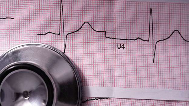
An Electrocardiogram, or ECG, is a fundamental test that records the electrical activity of the heart to detect abnormalities. It is a quick and painless method often used in routine cardiac evaluations.
- Symptoms/causes: Chest pain, palpitations, dizziness, or unexplained shortness of breath may prompt an ECG to identify arrhythmias or ischemia.
- Diagnosis: Electrodes placed on the chest capture electrical impulses that are displayed as waves on paper or digital format, highlighting irregularities.
- Treatment/management: ECG findings help guide further diagnostic testing and treatment plans including medication adjustment or lifestyle changes.
- What to expect: The procedure takes about 5-10 minutes and involves lying still while electrodes monitor heart function without discomfort.
This non-invasive test provides essential insight into heart health and helps clinicians make timely decisions for patient care.