
Dr Jitendra Singh
About Me
Dr Jitendra Singh
DO
Ophthalmologist
10 Years Experience
Biography
My Clinic
OPD Hours
| Monday | Morning | 10:00 AM | - | 02:00 PM |
| Evening | 06:00 PM | - | 09:00 PM | |
| Tuesday | Morning | 10:00 AM | - | 02:00 PM |
| Evening | 06:00 PM | - | 09:00 PM | |
| Wednesday | Morning | 10:00 AM | - | 02:00 PM |
| Evening | 06:00 PM | - | 09:00 PM | |
| Thursday | Morning | 10:00 AM | - | 02:00 PM |
| Evening | 06:00 PM | - | 09:00 PM | |
| Friday | Morning | 10:00 AM | - | 02:00 PM |
| Evening | 06:00 PM | - | 09:00 PM | |
| Saturday | Morning | 10:00 AM | - | 02:00 PM |
| Evening | 06:00 PM | - | 09:00 PM | |
| Sunday | Morning | 10:00 AM | - | 02:00 PM |
| Evening | 06:00 PM | - | 09:00 PM |
Treatments
Specialised care and treatments offered
ENDOPHTHALMITIS

An infection inside the eye called endophthalmitis presents rapidly after trauma or surgery. This condition represents a serious threat to sight needing urgent ophthalmologic treatment to prevent permanent vision loss.
- Identification & symptoms : Patients complain of worsening eye pain, redness increasing streaks from previous procedures , dramatic decrease in eyesight , sometimes eyelid swelling .
- Diagnostic tools : Clinical exam along with vitreous sampling under sterile conditions confirms infectious organisms enabling tailored antibiotic choice .
- Management plan : Intravitreal antibiotic injections combined with systemic therapy form mainstay treatment ; surgical vitrectomy removes infected debris if required .
- Prognostic information : Rapid intervention improves outcomes considerably although visual recovery depends on infection severity .
Understanding urgent nature alongside meticulous care offers best chance at salvaging vision from this rare but critical complication .
OPTIC NEURITIS
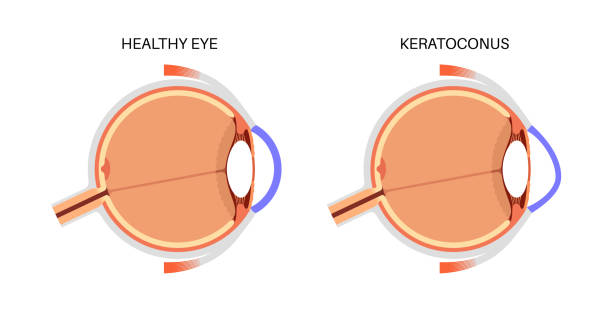
The inflammation of the optic nerve known as optic neuritis can cause sudden visual disturbances. It often affects young adults and may herald underlying neurological conditions such as multiple sclerosis (MS).
- Pertinent symptoms & causes:Sufferers usually experience painful eye movements accompanied by rapid onset of blurry vision or color desaturation in one eye; autoimmune reactions commonly underlie this condition.
- Eliciting diagnosis:MRI imaging alongside clinical assessment helps confirm inflammation of the optic nerve while excluding other causes like infections or tumors.
- Therapeutic approaches:Corticosteroid therapy expedites visual recovery though most patients improve spontaneously over weeks; further neurologic evaluation guides ongoing care if MS is suspected.
CORNEAL ULCER

A corneal ulcer is an open sore on the cornea typically caused by infection or injury. If untreated, it can lead to serious complications including vision loss or perforation of the eye wall.
- Main causes and signs: Commonly triggered by bacterial, fungal or viral infections; symptoms include redness, pain worsened by light exposure (photophobia), excessive tearing, discharge, and decreased vision.
- Diagnostic methods: Slit lamp examination paired with corneal staining highlights lesions; cultures may identify infectious agents for targeted therapy.
- Treatment modalities: Intensive antimicrobial drops are started promptly based on suspected cause; sometimes oral medications are added along with supportive care such as pain control.
- You should know:The healing process may be slow but adherence to therapy prevents progression into more serious outcomes such as scarring or perforation requiring surgery.
A timely medical evaluation ensures appropriate treatment heading off complications from these potentially sight-threatening infections.
RETINAL DETACHMENT
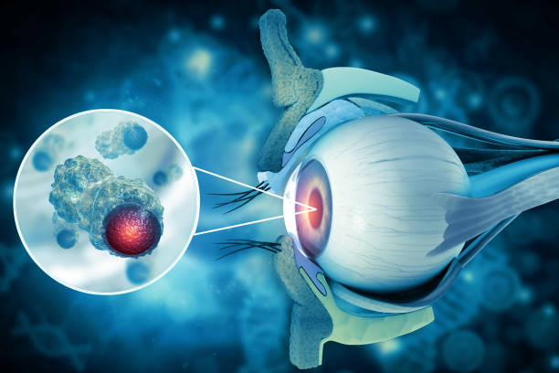
A retinal detachment occurs when the retina separates from its underlying supportive tissue. This threatens vision and usually demands urgent surgical repair to avoid permanent damage.
- Symptoms and causative factors: Patients often report sudden flashes of light, floaters or spots in vision, followed by a shadow or curtain effect over part of the visual field. It may be associated with trauma or high myopia.
- Diagnostic approach: Diagnosis relies on dilated fundus examination by an ophthalmologist using specialized equipment like an indirect ophthalmoscope.
- Treatment strategies: Surgical options include pneumatic retinopexy, scleral buckle placement, or vitrectomy depending on detachment severity and location.
- Outcome considerations: Early surgery greatly enhances chances for visual recovery; untreated detachment leads to irreversible blindness in affected areas.
This condition requires swift professional intervention as it constitutes a true ocular emergency demanding expert care.
CHEMICAL EYE BURNS
Chemical burns of the eye occur when hazardous substances come into contact with the ocular surface and can cause severe damage. These injuries require immediate action to reduce long-term complications.
- Causes and symptoms: Exposure typically involves acids or alkalis causing redness, pain, tearing, blurred vision, and swelling.
- Diagnosis: Clinical evaluation includes assessing extent of corneal injury and ocular surface involvement using fluorescein staining.
- Treatment and management: Immediate and copious irrigation with saline or water is vital. Follow-up care may include topical antibiotics, anti-inflammatory agents, and sometimes surgical interventions for severe burns.
- Prognosis expectations: Prompt first aid limits tissue damage; delayed treatment can result in scarring or vision impairment.
A rapid response following exposure plays a critical role in protecting vision after chemical injury to the eyes.
ACUTE ANGLE-CLOSURE GLAUCOMA
Acute angle-closure glaucoma is a sudden increase in intraocular pressure that can lead to vision loss if not promptly treated. This condition is an ocular emergency requiring immediate attention.
- Symptoms and causes: It often presents with severe eye pain, headache, blurred vision, halos around lights, nausea, and vomiting. It occurs when the drainage angle of the eye becomes blocked.
- Diagnosis: Eye pressure measurement (tonometry), slit-lamp examination, and gonioscopy confirm the diagnosis.
- Treatment options: Initial management includes medications to lower eye pressure quickly, such as topical beta-blockers or oral acetazolamide. Laser peripheral iridotomy is commonly performed to create an alternative drainage path.
- What to expect: Emergency treatment aims to prevent optic nerve damage; without prompt care, permanent vision loss can occur.
Timely intervention significantly improves prognosis, making awareness of symptoms crucial for preserving eyesight in this urgent condition.