
Dr Kumar Pravinchandra Doshi
About Me
Dr Kumar Pravinchandra Doshi
Cardiologist
Biography
My Clinic
OPD Hours
| Tuesday | Morning | 10:00 AM | - | 02:00 PM |
| Evening | 06:00 PM | - | 09:00 PM | |
| Wednesday | Morning | 10:00 AM | - | 02:00 PM |
| Evening | 06:00 PM | - | 09:00 PM | |
| Thursday | Morning | 10:00 AM | - | 02:00 PM |
| Evening | 06:00 PM | - | 09:00 PM | |
| Friday | Morning | 10:00 AM | - | 02:00 PM |
| Evening | 06:00 PM | - | 09:00 PM | |
| Saturday | Morning | 10:00 AM | - | 02:00 PM |
| Evening | 06:00 PM | - | 09:00 PM | |
| Sunday | Morning | 10:00 AM | - | 02:00 PM |
| Evening | 06:00 PM | - | 09:00 PM | |
| Thursday | Morning | 10:00 AM | - | 02:00 PM |
| Evening | 06:00 PM | - | 09:00 PM |
Treatments
Specialised care and treatments offered
STRESS TESTING

A stress test assesses how well your heart performs under physical exertion. It's essential for diagnosing conditions such as coronary artery disease or evaluating exercise tolerance post-cardiac events.
- Evaluation Criteria: Typically indicated if one experiences unexplained chest pains or has risk factors needing thorough evaluation; this helps ascertain safe exercise limits moving forward toward recovery plans
- Test Procedure: Patients usually walk on a treadmill while their heart rate and rhythm are monitored closely via ECG testing—an accurate depiction showing reactions under strain took place
- Potential Findings: Results indicate possible signs indicating blockages; abnormal responses suggest further investigation required through additional imaging modalities accordingly
- Recovery Insights: After performing exercise portion most participants have minimal side effects which resolve quickly! Detailed instructions afterward guide beneficial habits ensuring no complications arise later on Post-test follow-ups assist healthcare providers refining treatments based upon outcomes observed accurately
This method proves fruitful establishing effective therapies tailored towards individual needs focusing improving future prognoses through vigilant assessments applicable when necessary!
PERIPHERAL VASCULAR DISEASE MANAGEMENT
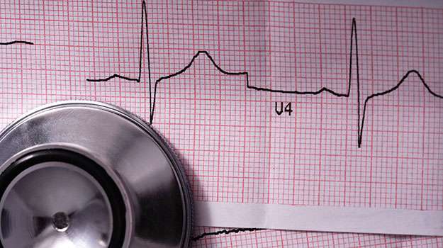
The management of peripheral vascular disease (PVD) focuses on improving blood circulation in patients with narrowed arteries outside of the coronary arteries. Effective management can significantly enhance quality of life for affected individuals.
- Causative Factors:The main causes include high cholesterol levels, diabetes, hypertension, and smoking-related damage to blood vessels leading to symptoms like leg cramps or pain during walking (claudication).
- Diagnosis Methodology: Diagnosis typically involves studies such as Doppler ultrasound or angiography which help visualize blood flow issues accurately.The physician evaluates risk factors profoundly before determining management steps
- Treatment Approaches: Management options range from lifestyle changes—such as diet modification—to medications enhancing blood flow; interventional options include angioplasty for severe cases.Classifying severity dictates consequent action appropriately
- Important Note: Early detection offers greater success rates! Regular follow-up appointments ensure sustained monitoring
Adequate management leads most patients towards symptom relief thereby reclaiming an active lifestyle!
TRANSESOPHAGEAL ECHOCARDIOGRAPHY (TEE)
This advanced imaging technique allows detailed visualization of the heart by inserting a small probe down the esophagus. TEE provides clearer images than standard echocardiograms due to proximity to cardiac structures.
- User Conditions: It is typically recommended when more information is needed about potential problems like valve disease or atrial fibrillation that cannot be seen adequately through conventional methods.
- Disease Detection: TEE can identify blood clots in the atrium and assess overall left ventricular function among other vital parameters while minimizing external interference from lung tissues or ribs.
- Please Note: Sedation may be required during the procedure; however, it is performed in an outpatient setting with monitored recovery times post-procedure being relatively quick.
This technique greatly enhances diagnostic capabilities in complex cases where traditional imaging techniques fall short!
HOLTER MONITOR
A Holter monitor is a portable device used for continuous monitoring of heart rhythms over 24-48 hours. It helps doctors evaluate irregularities that may not show up during standard exams.
- Purpose: This monitor is often prescribed if patients report palpitations or unusual symptoms suggesting arrhythmias.
- User-Friendly: Patients wear the device attached with electrodes that record heart activity throughout daily activities while maintaining normal routines.
- Diverse Interpretations: Data collected can reveal transient arrhythmias or correlate symptoms with specific activities or times of day.
- No Significant Side Effects: There are generally no side effects involved; some discomfort may arise from electrode placement but typically resolves quickly after removal.
The Holter monitor offers invaluable data for accurate diagnosis and informed treatment decisions regarding cardiac health issues.
ECHOCARDIOGRAPHY
Echocardiography is a diagnostic tool that uses sound waves to create detailed images of the heart's structure and function. This non-invasive test plays a crucial role in diagnosing various cardiac conditions without requiring any surgery.
- Symptoms: Patients may undergo this test when experiencing unexplained shortness of breath, palpitations, or chest pain.
- How it Works: Sound waves create echoes as they bounce off heart structures. These echoes are then converted into images by a computer for analysis.
- Diagnostic Uses: Echocardiograms help evaluate heart valve performance, assess chamber sizes, and check for fluid around the heart.
- No Risks Involved: Being non-invasive means there are minimal risks associated with echocardiography; patients can usually return to their daily routine immediately after the exam.
This versatile tool provides important insights into cardiac health and guides treatment planning effectively.
CORONARY ANGIOPLASTY
Coronary angioplasty is a minimally invasive procedure used to open blocked coronary arteries, allowing for improved blood flow to the heart muscle. This technique is commonly employed in patients with coronary artery disease, leading to better heart health and function.
- Symptoms: Patients often experience chest pain (angina), shortness of breath, or fatigue during physical activity due to reduced blood flow.
- Diagnosis: Diagnosis typically involves an electrocardiogram (ECG), echocardiogram, and stress tests, followed by angiography to visualize blockages.
- Treatment Options: The primary intervention involves placing a balloon at the site of blockage, which is inflated to widen the artery. Often, a stent may be placed to keep the artery open.
- Recovery Expectation: Many patients can return home within hours and resume normal activities in a few days, although some precautions are necessary.
This procedure has been shown to significantly reduce symptoms and improve quality of life for individuals suffering from chest pain or related heart conditions.