
Dr Malik
About Me
Dr Malik
Cardiologist
Biography
My Clinic
OPD Hours
| Tuesday | Morning | 10:00 AM | - | 02:00 PM |
| Evening | 06:00 PM | - | 09:00 PM | |
| Wednesday | Morning | 10:00 AM | - | 02:00 PM |
| Evening | 06:00 PM | - | 09:00 PM | |
| Thursday | Morning | 10:00 AM | - | 02:00 PM |
| Evening | 06:00 PM | - | 09:00 PM | |
| Friday | Morning | 10:00 AM | - | 02:00 PM |
| Evening | 06:00 PM | - | 09:00 PM | |
| Saturday | Morning | 10:00 AM | - | 02:00 PM |
| Evening | 06:00 PM | - | 09:00 PM | |
| Sunday | Morning | 10:00 AM | - | 02:00 PM |
| Evening | 06:00 PM | - | 09:00 PM | |
| Thursday | Morning | 10:00 AM | - | 02:00 PM |
| Evening | 06:00 PM | - | 09:00 PM |
Treatments
Specialised care and treatments offered
CORONARY ANGIOGRAPHY

This specialized imaging technique utilizes X-rays to visualize coronary arteries after injecting contrast dye into bloodstream providing real-time insights into myocardial perfusion status helping identify obstructive lesions..
- Symptoms Necessitating Testing: Chest pain along with risk factors such as hypertension hyperlipidemia obesity confirms eligibility requiring assessment underlying factors compromising hemodynamics occurring inside circulation.
- Diagnostic Methodology & Procedure: Catheters are precisely manipulated towards coronary vessels delivering contrast enabling visualization captured via fluoroscopy monitored continuously throughout session lasting approximately one hour ensuring minimal inconvenience..
- Outcomes Guiding Therapies: Maintenance strategies may include drug therapy lifestyle modification shunt insertions leading up preventive interventions aimed alleviating anginal episodes post evaluation results obtained considered respective patients condition suitability underwent examination. .
CARDIAC CATHETERIZATION
Cardiac catheterization involves threading a thin tube through blood vessels into the heart for diagnostic purposes. It's pivotal for evaluating coronary artery disease and measuring pressures within heart chambers more intricately than external tests allow.
STRESS TESTING
A stress test measures how well your heart functions under physical stress. It helps determine if there are any exercise-induced arrhythmias or blockages within coronary arteries affecting blood flow during increased exertion levels.
- Symptoms Indicating Need: Common indications include unexplained shortness of breath or chest discomfort when engaging in physical activity.
- Diagnostic Methods: Patients typically walk on a treadmill while their electrocardiogram and vital signs are monitored closely throughout different stages of increasing difficulty.
- Treatment Decisions: Results can guide treatment options ranging from lifestyle modifications to surgical intervention depending on detected cardiovascular issues under strain conditions.
HOLTER MONITOR
A Holter monitor is a portable device that continuously records the heart's rhythms for 24 hours or longer. It helps detect irregularities in heartbeat patterns that might not be caught during a standard ECG.
- Symptoms: Patients with chest discomfort, fainting spells, or arrhythmia symptoms often undergo Holter monitoring for diagnosis.
- Diagnosis Process: Electrodes are attached to your chest and connected to the Holter monitor worn throughout daily activities. You will also keep a diary of symptoms during this period.
- Treatment Options: Based on results from this monitoring period, doctors can recommend appropriate treatments such as medication adjustments or further diagnostic testing.
- What to Expect: The device is lightweight and worn comfortably under clothing while you perform normal activities; it usually yields comprehensive data about your heart's functioning during daily life.
This dynamic approach allows healthcare providers to understand how everyday activities influence heart health better than static tests alone.
ECHOCARDIOGRAPHY
Echocardiography is an imaging technique that uses ultrasound waves to create detailed images of the heart's structure and function. It enables cardiologists to visualize how well the heart is pumping blood and identify any structural abnormalities.
- Symptoms: Common reasons for an echocardiogram include unexplained fatigue, heart murmurs, or signs of heart failure.
- Diagnosis: This test is performed using a transducer placed on the chest, which sends ultrasound waves that echo back. This information is then converted into visual images on a monitor.
- Treatment Management: Depending on findings from the echocardiogram, treatment may involve lifestyle changes, medications, or referrals for surgical interventions if needed.
- What to Expect: Patients typically lie on their left side during the test, which lasts about 30-60 minutes. It is non-invasive and poses no risks.
Overall, echocardiography provides valuable information regarding cardiac function essential for tailored patient care.
ELECTROCARDIOGRAM (ECG)
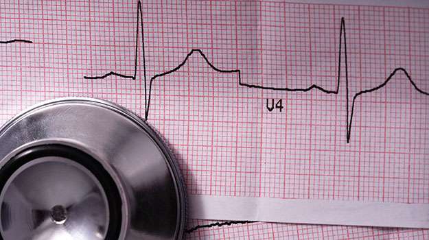
An Electrocardiogram (ECG) is a non-invasive test that records the electrical activity of the heart over a period of time. This test helps in diagnosing various heart conditions by providing insights into the heart's rhythm and any abnormalities present.
- Symptoms: Patients may experience chest pain, palpitations, dizziness, or shortness of breath prompting this test.
- Diagnosis: An ECG is typically performed in a doctor's office or hospital setting. Electrodes are attached to the skin, allowing for real-time monitoring of heart activity.
- Treatment Options: While an ECG itself does not treat conditions, it aids in determining subsequent management plans like medications or further testing.
- What to Expect: The procedure is painless and takes only a few minutes. You will be asked to lie still while the electrodes record your heart's activity.
This diagnostic tool plays a crucial role in assessing heart health and guiding further investigations if necessary.