
Dr Manish Sharma
About Me
Dr Manish Sharma
DO
Ophthalmologist
10 Years Experience
Biography
My Clinic
OPD Hours
| Monday | Morning | 10:00 AM | - | 02:00 PM |
| Evening | 06:00 PM | - | 09:00 PM | |
| Tuesday | Morning | 10:00 AM | - | 02:00 PM |
| Evening | 06:00 PM | - | 09:00 PM | |
| Wednesday | Morning | 10:00 AM | - | 02:00 PM |
| Evening | 06:00 PM | - | 09:00 PM | |
| Thursday | Morning | 10:00 AM | - | 02:00 PM |
| Evening | 06:00 PM | - | 09:00 PM | |
| Friday | Morning | 10:00 AM | - | 02:00 PM |
| Evening | 06:00 PM | - | 09:00 PM | |
| Saturday | Morning | 10:00 AM | - | 02:00 PM |
| Evening | 06:00 PM | - | 09:00 PM | |
| Sunday | Morning | 10:00 AM | - | 02:00 PM |
| Evening | 06:00 PM | - | 09:00 PM |
Treatments
Specialised care and treatments offered
DIABETIC RETINOPATHY LASER TREATMENT
This condition arises from damage to retinal blood vessels due to prolonged high blood sugar in people with diabetes. Timely laser therapy helps prevent progression towards blindness.
- Diabetic retinopathy usually starts asymptomatic but may progress causing floaters, blurred/darkened central or peripheral vision.
- Diagnosis requires detailed retinal examination often supplemented by fluorescein angiography which highlights leaking microaneurysms/focuses ischemic zones.
- Panretinal photocoagulation laser treatment targets peripheral areas reducing abnormal vessel growth while focal laser treats localized macular edema.
- The procedure preserves remaining eyesight preventing more severe consequences like vitreous hemorrhage or tractional retinal detachment.
- Follow-up diabetic control plus periodic ophthalmic exams essential post-treatment.
Laser treatments remain fundamental in managing diabetic retinopathy effectively even now alongside advancing pharmacotherapy making early diagnosis critical.
RETINAL DETACHMENT REPAIR
A retinal detachment occurs when the thin neurosensory retina separates from its underlying supportive tissue, risking permanent vision loss without emergency intervention. Surgical repair aims to reattach the retina promptly to preserve sight.
- Symptoms commonly include sudden appearance of floaters, flashes of light, shadow/curtain over part of vision, or reduced visual acuity in one eye.
- Diagnosis involves detailed dilated fundus examination with possible ultrasonography if view is obscured by vitreous hemorrhage or other media opacity.
- Treatment options vary based on severity: pneumatic retinopexy injects gas bubble into vitreous; scleral buckling places silicone bands around eyeball; vitrectomy removes vitreous gel allowing retinal flattening via internal tamponade techniques using gas or silicone oil;
CORNEAL TRANSPLANTATION (KERATOPLASTY)
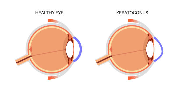
A damaged or diseased cornea that causes significant vision impairment may require surgical replacement through corneal transplantation. This restores corneal clarity by grafting donor tissue onto the patient's eye surface.
- Indications include advanced corneal scars, keratoconus, dystrophies, or infections unresponsive to medical treatment.
- An extensive preoperative assessment evaluates ocular surface health and maps corneal dimensions for graft selection.
- The transplant can be full-thickness (penetrating keratoplasty) or partial-thickness (lamellar keratoplasty) depending on disease extent.
- Surgical success depends on meticulous technique and postoperative anti-rejection therapy using corticosteroids and immunosuppressants if needed.
- The recovery timeline varies but requires regular follow-up visits for months to monitor graft clarity and address complications such as infection or rejection episodes promptly.
This surgery offers hope for restoring functional vision in eyes with previously irreversible corneal damage when appropriately indicated and managed diligently afterward.
GLAUCOMA DRAINAGE IMPLANT SURGERY
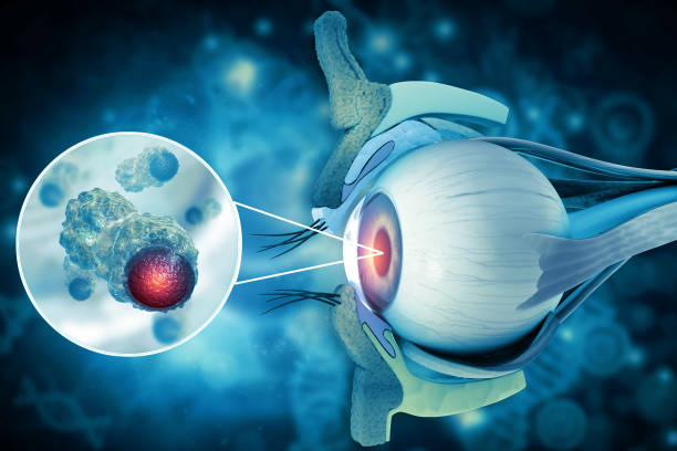
Glaucoma drainage implant surgery helps lower intraocular pressure in patients where medications or conventional surgeries are insufficient. This can prevent further damage to the optic nerve responsible for vision.
- The condition is characterized by increased pressure inside the eye leading to gradual peripheral vision loss.
- Diagnosis involves measuring intraocular pressure, visual field testing, and optic nerve imaging.
- The procedure inserts a small tube or plate device that facilitates drainage of aqueous humor from inside the eye to reduce pressure effectively.
- Surgery is usually reserved for advanced cases or those who have had prior unsuccessful glaucoma treatments.
- Post-surgical monitoring ensures proper function of the implant and management of any complications such as infection or hypotony (low eye pressure).
This intervention can be sight-saving for patients facing progressive glaucomatous damage despite other therapies.
LASIK SURGERY
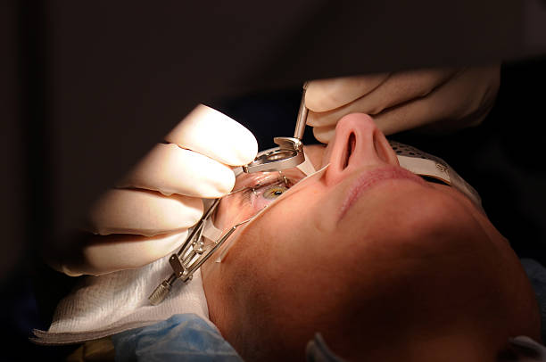
Laser-Assisted In Situ Keratomileusis (LASIK) corrects refractive errors to reduce dependence on glasses or contact lenses. It reshapes the cornea to improve how light is focused onto the retina.
- Common indications include myopia, hyperopia, and astigmatism causing blurry vision.
- A thorough preoperative evaluation assesses corneal thickness, pupil size, and overall eye health to ensure suitability.
- The procedure uses an excimer laser to create a precise corneal flap before remodeling the underlying tissue.
- Patients usually experience improved vision within 24 hours with minimal discomfort.
- Follow-up visits monitor healing; some may require enhancements for optimal results.
LASIK offers rapid recovery with few complications when performed by skilled specialists using modern technology.
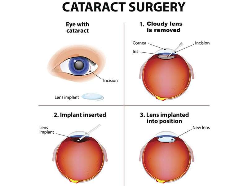
CATARACT SURGERY
Cataracts cause clouding of the eye's natural lens, leading to decreased vision that can affect daily activities. Surgical removal of the cataract and replacement with an artificial lens is a common and highly effective treatment.
- Symptoms include blurred vision, glare, difficulty seeing at night, and faded colors.
- Diagnosis is confirmed through a comprehensive eye exam including visual acuity tests and slit-lamp examination.
- Treatment involves phacoemulsification or ultrasound-assisted removal of the cloudy lens followed by implantation of an intraocular lens (IOL).
- The procedure is typically outpatient with local anesthesia and rapid visual recovery.
- Postoperative care includes use of prescribed eye drops and avoiding strenuous activity for a short period.
This surgery often significantly improves vision and quality of life, with low risk when performed by an experienced ophthalmic surgeon.