
Dr Mukul Goyal
About Me
Dr Mukul Goyal
MD
Oncologist
10 Years Experience
Biography
My Clinic
OPD Hours
| Monday | Morning | 10:00 AM | - | 02:00 PM |
| Evening | 06:00 PM | - | 09:00 PM | |
| Tuesday | Morning | 10:00 AM | - | 02:00 PM |
| Evening | 06:00 PM | - | 09:00 PM | |
| Wednesday | Morning | 10:00 AM | - | 02:00 PM |
| Evening | 06:00 PM | - | 09:00 PM | |
| Thursday | Morning | 10:00 AM | - | 02:00 PM |
| Evening | 06:00 PM | - | 09:00 PM | |
| Friday | Morning | 10:00 AM | - | 02:00 PM |
| Evening | 06:00 PM | - | 09:00 PM | |
| Saturday | Morning | 10:00 AM | - | 02:00 PM |
| Evening | 06:00 PM | - | 09:00 PM | |
| Sunday | Morning | 10:00 AM | - | 02:00 PM |
| Evening | 06:00 PM | - | 09:00 PM |
Treatments
Specialised care and treatments offered
PET SCAN FOR CANCER DETECTION
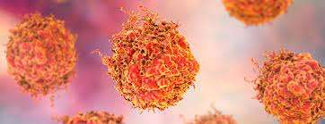
A PET (Positron Emission Tomography) scan is an advanced imaging technique that helps highlight functioning areas within tissues using radioactive tracers injected into the bloodstream. This method is powerful in detecting active cancer cells compared to surrounding healthy tissues!
- Symptoms Prompting Assessment: Weight loss without clear cause along severe fatigue could lead physicians recommending this important diagnostic tool!
- Process Overview: After tracer administration via IV route patients generally wait prior scanning—a specialized camera detects emitted signals highlighting regions exhibiting abnormal metabolic activity indicative of rapid growth typical of malignant lesions!
- Treatment Application: PET scans assist providers defining accurate staging influencing decisions concerning localized surgical approaches versus larger multisite systemic therapies thereby aiding prognosis disclosures robustly!
- Your Experience Level: Overall encounter lasts between two hours including preparation time—patients should feel reasonably comfortable throughout procedure aside occasional sensations from intravenous lines utilized administratively!
Emphasizing its pivotal role reflecting physiological behavior enhances overall investigative strategies substantially benefiting patients' comprehensive care narratives down pathway towards optimal outcomes.
BLOOD TESTS FOR TUMOR MARKERS
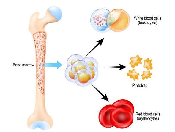
BLOOD TESTS SPECIFIC FOR TUMOR MARKERS ARE ESSENTIAL IN ONCOLOGY FOR MONITORING AND DIAGNOSIS OF CANCER. These tests measure specific substances produced by cancer cells that can be detected in blood samples and play an important role in both diagnosis and treatment monitoring over time.
- Key Indicators: Common tumor markers include PSA for prostate cancer and CA-125 primarily associated with ovarian cancer.
- Diagnostic Method: Blood samples are sent to laboratories where assays identify elevated levels of these markers; however, they are not solely definitive.
- Treatment Implications: Depending on results indicating rising marker levels post-treatment may suggest recurrence necessitating further investigation; conversely stable levels indicate ongoing successful management under care plan directives.
- Patient Experience: Patients are recommended regular testing intervals which ease anxieties related regarding possible progression allowing appropriate adjustments per findings.
Monitoring tumor markers become foundational elements seeking optimal outcomes throughout cancer trajectory with insights guiding interventions effectively!
ENDOSCOPIC ULTRASOUND (EUS)

An endoscopic ultrasound (EUS) is an advanced imaging technique that combines endoscopy with ultrasound technology. This procedure allows healthcare providers to obtain high-resolution images of internal organs while also allowing tissue sampling through fine-needle aspiration if abnormalities are found.
- Sparking Symptoms: EUS may be indicated if symptoms like persistent abdominal pain or difficulty swallowing arise, possibly indicating gastrointestinal cancers.
- The Procedure Explained: A thin tube with an ultrasound probe is inserted through the mouth or rectum, producing detailed images of nearby structures as well as enabling biopsies when necessary.
- Cancer Management: It helps establish early diagnoses and contributes valuable information for staging cancers affecting digestive organs with unmatched detail.
- Your Expectations:This minimally invasive procedure usually lasts around 30-60 minutes; discomfort may occur during sedation recovery but generally resolves quickly
EUS plays a crucial role in developing personalized treatment approaches based on precise anatomy assessment.
MRI FOR TUMOR EVALUATION
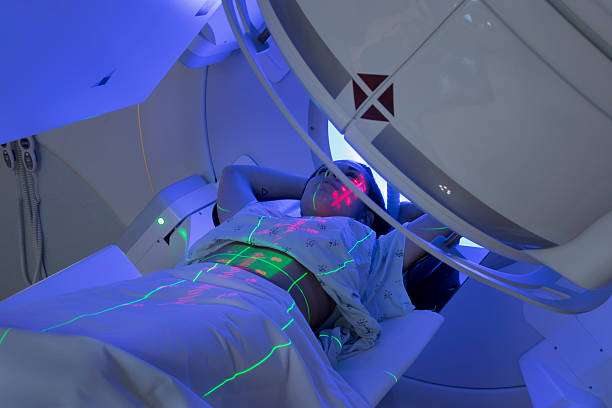
An MRI (Magnetic Resonance Imaging) scan is a powerful diagnostic tool used to visualize soft tissue structures within the body. For oncology patients, it provides critical information about tumor size, location, and potential involvement with surrounding tissues.
- Main Purpose: MRIs are particularly useful for assessing brain tumors and cancers in organs like the liver or prostate.
- Disease Detection: This imaging method does not use ionizing radiation; instead, it employs magnetic fields and radio waves to produce high-resolution images.
- Treatment Insights: The clarity of MRI images aids oncologists in determining suitable therapeutic approaches based on tumor characteristics.
- Your Experience: Patients should expect to remain still within a tube-like scanner during the process; some may experience claustrophobia but can ask for assistance if needed.
The detailed images obtained allow for informed discussions about treatment options following evaluation.
CT SCAN FOR CANCER STAGING
A CT scan is an imaging technique that provides detailed cross-sectional images of the body's organs and tissues. It plays a crucial role in cancer staging and helps oncologists assess the extent of disease spread within the body.
- Symptoms Indicating Need: Symptoms such as unexplained weight loss or persistent pain may prompt a CT scan to evaluate potential underlying conditions.
- Diagnosis Process: This non-invasive test uses X-rays and computer technology to create detailed images after injecting a contrast dye for better visibility of tumors.
- Treatment Planning: Results from the scan inform necessary treatment plans, whether it involves surgery or chemotherapy.
- What to Expect: The scan typically takes 30 minutes to an hour, with patients advised to lie still while images are captured. There might be minor side effects from the contrast material used.
The results will help guide your oncologist on the most effective treatment strategies moving forward.
BREAST BIOPSY
A breast biopsy is a medical procedure that involves the removal of cells or tissues from the breast to determine if cancer is present. This procedure is vital for an accurate diagnosis and is typically recommended when there are abnormalities detected in mammograms or physical examinations.
- Symptoms: Common signs prompting a biopsy include lumps, changes in breast shape, or unusual discharge.
- Diagnostic Method: A biopsy can be performed using various techniques such as fine-needle aspiration or excisional biopsy, often guided by imaging tests.
- Treatment Options: If cancer is diagnosed, treatment may involve surgery, radiation therapy, or systemic therapies like chemotherapy.
- Expectations: Patients can expect some discomfort during the procedure but typically recover quickly. Results usually take a few days to be processed and reviewed.
This procedure is essential for guiding further treatment decisions.