
Dr Narendra Sing
About Me
Dr Narendra Sing
DO
Ophthalmologist
10 Years Experience
Biography
My Clinic
OPD Hours
| Monday | Morning | 10:00 AM | - | 02:00 PM |
| Evening | 06:00 PM | - | 09:00 PM | |
| Tuesday | Morning | 10:00 AM | - | 02:00 PM |
| Evening | 06:00 PM | - | 09:00 PM | |
| Wednesday | Morning | 10:00 AM | - | 02:00 PM |
| Evening | 06:00 PM | - | 09:00 PM | |
| Thursday | Morning | 10:00 AM | - | 02:00 PM |
| Evening | 06:00 PM | - | 09:00 PM | |
| Friday | Morning | 10:00 AM | - | 02:00 PM |
| Evening | 06:00 PM | - | 09:00 PM | |
| Saturday | Morning | 10:00 AM | - | 02:00 PM |
| Evening | 06:00 PM | - | 09:00 PM | |
| Sunday | Morning | 10:00 AM | - | 02:00 PM |
| Evening | 06:00 PM | - | 09:00 PM |
Treatments
Specialised care and treatments offered
FUNDUS PHOTOGRAPHY
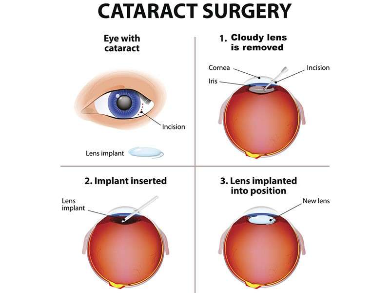
This procedure involves capturing detailed images of the interior surface of the eye (fundus), including retina, optic disc, macula and posterior pole. It aids documentation and diagnosis of various ocular diseases.
- Symptoms/causes: Indicated when there is suspicion of diabetic retinopathy changes , macular degeneration , optic nerve abnormalities , or unexplained visual disturbances .
- Diagnosis: Specialized cameras take color pictures after pupil dilation providing permanent records for comparison over time .
- Treatment/management: Images help ophthalmologists monitor disease progression , plan interventions like laser photocoagulation , and educate patients about their condition .
- What to expect: Non-invasive , painless imaging session usually lasting less than 10 minutes with brief use of dilating drops .
Fundus photography enhances understanding of ocular health status enabling precise follow-up strategies tailored per individual needs .
VISUAL FIELD TESTING
This test evaluates peripheral (side) vision which can be affected by diseases such as glaucoma or neurological disorders. It helps identify blind spots invisible during routine exams.
- Symptoms/causes: Advised if there are concerns about unexplained vision loss or during routine glaucoma monitoring visits.
- Diagnosis: Performed using automated devices where patients respond to lights appearing in different areas inside a dome-shaped instrument. Treatment/management: Anomalies detected guide further diagnostic workup and tailored therapeutic decisions aimed at halting progression of underlying disease processes.
- What to expect: The test requires patient cooperation over several minutes; results produce maps highlighting areas with decreased sensitivity.
Regular visual field assessments play an important role in comprehensive eye care aiming at preserving functional sight over time.
TONOMETRY FOR INTRAOCULAR PRESSURE MEASUREMENT
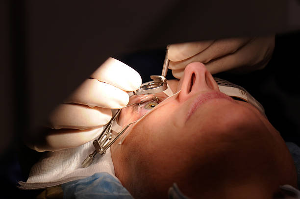
The measurement of intraocular pressure (IOP) plays a critical role in glaucoma screening and management. Elevated pressure might damage the optic nerve leading to vision loss if untreated.
- Symptoms/causes: Typically performed on individuals at risk or those experiencing symptoms like gradual peripheral vision loss.
- Diagnosis: Several methods exist including puff air tests or contact-based applanation tonometry under topical anesthesia.
- Treatment/management: Elevated IOP may require medication adherence, lifestyle changes, laser therapy, or surgery based on severity and response to treatment.
- What to expect: The procedure is quick with minimal discomfort; some methods avoid direct contact while others use gentle corneal touch after numbing drops are applied.
This screening assists in early detection preventing potential irreversible optic nerve damage through timely intervention strategies.
SLIT LAMP EXAMINATION
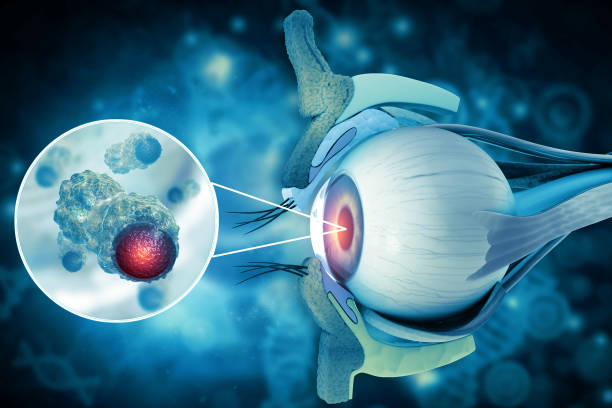
A slit lamp exam offers magnified visualization of the front structures of the eye including eyelids, cornea, conjunctiva, iris, and lens. It helps pinpoint abnormalities affecting these areas.
- Symptoms/causes: Used when patients report irritation, redness, pain, or visual changes suggesting surface or anterior segment issues.
- Diagnosis: The device combines bright light and magnification allowing detailed inspection by the ophthalmologist.
- Treatment/management: Findings can lead to prescribing medications for infections/inflammations or planning surgical interventions if necessary.
- What to expect: The exam is non-invasive but may involve instilling eye drops to dilate pupils for better visualization depending on clinical need.
This examination method is fundamental in detecting a broad spectrum of ocular conditions quickly and accurately.
OPTICAL COHERENCE TOMOGRAPHY (OCT)
This imaging technique provides high-resolution cross-sectional views of the retina, allowing detailed evaluation of its layers. It's crucial for diagnosing various retinal disorders.
- Symptoms/causes: Recommended for patients with symptoms like distorted vision or known retinal diseases including macular degeneration or diabetic retinopathy.
- Diagnosis: OCT uses light waves to capture detailed images without any discomfort or radiation exposure.
- Treatment/management: Results guide treatment plans such as injections, laser therapy, or monitoring progression over time.
- What to expect: The scan is quick and painless; you rest your chin on a support while the device captures images within minutes.
This test is invaluable in providing clear insight into retinal health and aids in personalized treatment approaches.
COMPREHENSIVE EYE EXAMINATION
A thorough eye exam is essential for maintaining good vision and overall eye health. This detailed assessment helps detect a variety of eye conditions early, even before symptoms appear.
- Symptoms/causes: Often performed routinely or when experiencing blurred vision, eye discomfort, or headache.
- Diagnosis: Includes tests such as visual acuity measurement, refraction assessment, pupil response check, and intraocular pressure measurement.
- Treatment/management: Based on findings, recommendations may include prescription glasses, further specialized testing, or referral to treatment options.
- What to expect: You will experience a non-invasive procedure that may take 20-45 minutes covering multiple steps to evaluate different parts of the eye.
This examination is foundational for detecting early signs of diseases and ensuring timely management to preserve vision.