
Dr Navneet Mehta
About Me
Dr Navneet Mehta
MD
Cardiologist
10 Years Experience
Biography
My Clinic
OPD Hours
| Monday | Morning | 10:00 AM | - | 02:00 PM |
| Evening | 06:00 PM | - | 09:00 PM | |
| Tuesday | Morning | 10:00 AM | - | 02:00 PM |
| Evening | 06:00 PM | - | 09:00 PM | |
| Wednesday | Morning | 10:00 AM | - | 02:00 PM |
| Evening | 06:00 PM | - | 09:00 PM | |
| Thursday | Morning | 10:00 AM | - | 02:00 PM |
| Evening | 06:00 PM | - | 09:00 PM | |
| Friday | Morning | 10:00 AM | - | 02:00 PM |
| Evening | 06:00 PM | - | 09:00 PM | |
| Saturday | Morning | 10:00 AM | - | 02:00 PM |
| Evening | 06:00 PM | - | 09:00 PM | |
| Sunday | Morning | 10:00 AM | - | 02:00 PM |
| Evening | 06:00 PM | - | 09:00 PM |
Treatments
Specialised care and treatments offered
BLOOD PRESSURE MONITORING

Blood pressure monitoring is fundamental in assessing cardiovascular risk factors and guiding hypertension treatment plans essential for preventing stroke and heart failure complications over time.
CORONARY ANGIOGRAPHY
This invasive imaging technique visualizes the coronary arteries supplying blood to the heart muscle. It helps identify narrowing or blockages responsible for angina and myocardial infarction risks requiring intervention.
- Symptoms/causes: Indicated when non-invasive tests suggest significant coronary artery disease presence causing chest pain or other ischemic symptoms.
- Diagnosis: A catheter inserted usually through wrist or groin delivers contrast dye into coronary vessels visible under X-ray fluoroscopy allowing precise localization of blockages.
HOLTER MONITORING
The Holter monitor is a portable device worn over 24-48 hours that continuously records your heart’s electrical activity. It helps detect intermittent arrhythmias not captured during standard ECGs done in clinics.
- Symptoms/causes: Used when patients report episodic palpitations, fainting spells, dizziness or unexplained fatigue without abnormal findings in initial ECGs.
- Diagnosis: Electrodes attached to the chest connect to a compact recorder tracking every heartbeat over extended durations during normal daily activities and sleep.
- Treatment/management: Data collected identifies abnormal rhythms requiring medication adjustment or advanced therapies such as pacemaker implantation if necessary.
- What to expect: Patients can carry out routine activities but must avoid water exposure; device removal occurs after monitoring period ends for data analysis by physicians.
This ambulatory monitoring offers valuable insights ensuring accurate detection of elusive cardiac irregularities impacting quality of life and safety.
STRESS TEST (TREADMILL TEST)
A Stress Test evaluates how well your heart functions during physical exertion. It is essential for identifying coronary artery disease and exercise-induced arrhythmias that may not be apparent at rest.
- Symptoms/causes: Recommended for patients presenting with chest discomfort on exertion or irregular heartbeat sensations during physical activity.
- Diagnosis: The patient walks on a treadmill or pedals a stationary bike while heart rate, rhythm, blood pressure, and ECG are closely monitored continuously.
- Treatment/management: Test outcomes assist cardiologists in deciding if further diagnostic procedures like angiography are warranted or optimizing medical management.
- What to expect: The procedure is generally safe but involves mild exertion; monitoring continues post-exercise until vital signs normalize completely.
This dynamic assessment effectively reveals hidden cardiac problems triggered by stress that resting tests might miss.
ECHOCARDIOGRAPHY
Echocardiography uses ultrasound waves to create moving images of the heart. It allows visualization of heart chambers, valves, and blood flow patterns to evaluate cardiac function comprehensively.
- Symptoms/causes: Indicated for detecting valve diseases, cardiomyopathies, pericardial effusions, or congenital defects causing symptoms such as fatigue and breathlessness.
- Diagnosis: A transducer placed on the chest emits sound waves that reflect off cardiac structures producing detailed images interpreted by specialists.
- Treatment/management: Results help determine disease severity and guide medical or surgical interventions depending on findings like valve abnormalities or pumping efficiency.
- What to expect: This non-invasive procedure usually lasts around 30-60 minutes without discomfort and has no radiation exposure risk.
Echocardiography remains an indispensable diagnostic modality providing critical insights into cardiac anatomy and functionality in various clinical scenarios.
ELECTROCARDIOGRAM (ECG)
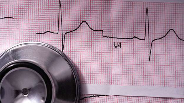
An Electrocardiogram, commonly known as ECG, is a vital tool for assessing the electrical activity of the heart. It helps to detect abnormalities in heart rhythm and structure by recording the heart’s electrical signals.
- Symptoms/causes: Often performed when patients experience chest pain, palpitations, dizziness, or unexplained shortness of breath.
- Diagnosis: The procedure involves placing electrodes on the skin to capture electrical impulses generated by the heart.
- Treatment/management: The ECG results guide physicians in diagnosing arrhythmias, ischemia, or myocardial infarction and determining appropriate treatment strategies.
- What to expect: The test is painless, non-invasive, and takes just a few minutes with immediate results available for evaluation.
This simple yet crucial exam is often the first step towards understanding heart health and planning further investigation if required.