
Dr O P Agrawal P
About Me
Dr O P Agrawal P
MS
Orthopaedic Surgeons
10 Years Experience
Biography
My Clinic
OPD Hours
| Monday | Morning | 10:00 AM | - | 02:00 PM |
| Evening | 06:00 PM | - | 09:00 PM | |
| Tuesday | Morning | 10:00 AM | - | 02:00 PM |
| Evening | 06:00 PM | - | 09:00 PM | |
| Wednesday | Morning | 10:00 AM | - | 02:00 PM |
| Evening | 06:00 PM | - | 09:00 PM | |
| Thursday | Morning | 10:00 AM | - | 02:00 PM |
| Evening | 06:00 PM | - | 09:00 PM | |
| Friday | Morning | 10:00 AM | - | 02:00 PM |
| Evening | 06:00 PM | - | 09:00 PM | |
| Saturday | Morning | 10:00 AM | - | 02:00 PM |
| Evening | 06:00 PM | - | 09:00 PM | |
| Sunday | Morning | 10:00 AM | - | 02:00 PM |
| Evening | 06:00 PM | - | 09:00 PM |
Treatments
Specialised care and treatments offered
X-RAYS
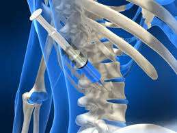
X-rays are fundamental imaging tests widely used to visualize bones and detect fractures. They provide quick diagnostic insights required for assessing orthopedic injuries efficiently.
- This method is primarily indicated when individuals present with acute pain or swelling, suggesting potential fractures due to trauma or falls occurring shortly before examination.
ELECTROMYOGRAPHY (EMG)
Electromyography (EMG) evaluates muscle function and nerve conduction using electrical activity recordings. This test helps diagnose neuromuscular disorders affecting muscles' ability to receive signals from nerves effectively.
- Common symptoms necessitating an EMG include muscle weakness, cramps, twitching or numbness that may suggest nerve issues affecting muscle control.
- The procedure involves placing small electrodes on targeted muscles while they contract gently; this records electrical signals which are analyzed subsequently by specialists.
An EMG provides valuable insights into muscular health and nerve integrity that can direct suitable treatment strategies based on findings observed during testing.
CT SCAN OF THE SPINE

A CT scan (Computed Tomography) of the spine provides comprehensive images of vertebrae and surrounding tissues, aiding in diagnosing spinal disorders. This cross-sectional imaging technique captures fine details not always visible on regular X-rays.
- This advanced imaging is indicated when patients experience chronic back pain, weakness in limbs, or following traumatic injuries to assess structural damage thoroughly.
- The process begins with positioning the patient on a scanner bed while cross-sectional images are taken at multiple angles to create 3D representations of the spine's anatomy.
- Your healthcare provider will review the findings to identify conditions like herniated discs, spinal stenosis, or tumors requiring attention.
A CT scan serves as an essential diagnostic tool for formulating appropriate management plans based on detailed visual information about spinal health.
BONE DENSITY TEST

A bone density test measures the amount of mineral content in bones and assesses their strength. This assessment is essential for diagnosing osteoporosis and determining fracture risk.
- The primary reason for undergoing this test includes sudden fractures, especially in older adults or those with specific risk factors like prolonged steroid use.
- Methods like Dual-Energy X-ray Absorptiometry (DEXA) scans are commonly employed for accurate measurements.
- If low bone density is detected, management strategies may involve lifestyle modifications along with medications to strengthen bone health.
- This test helps establish a baseline for monitoring bone health over time, allowing timely interventions when necessary.
Understanding bone density plays a critical role in preventing fractures and maintaining long-term skeletal health.
MRI OF THE KNEE

An MRI (Magnetic Resonance Imaging) scan of the knee is a crucial diagnostic tool that provides detailed images of soft tissues around the knee joint. This imaging technique is non-invasive and does not involve radiation.
- The procedure is commonly indicated for evaluating suspicion of tears in ligaments or cartilage due to injury.
- Symptoms leading to an MRI may include persistent swelling, pain during movement, or restricted range of motion.
- A radiologist interprets the images obtained from the MRI to identify abnormalities such as meniscus tears or ligament injuries.
- This imaging modality can assist in planning further management options including surgical intervention if necessary.
An MRI offers invaluable insights into knee conditions that might not be visible on standard X-rays, guiding effective treatment strategies.
ARTHROSCOPY

Arthroscopy is a minimally invasive surgical procedure used to diagnose and treat joint problems. It involves inserting a thin tube with a camera into the joint space, providing real-time visuals for evaluation and intervention.
- Common indications for arthroscopy include persistent joint pain, swelling, or limited mobility that has not responded to conservative measures.
- Diagnosis is typically confirmed through imaging studies such as X-rays or MRI, followed by the arthroscopy procedure itself.
- Treatment options may include cleaning out damaged tissue, repairing ligaments, or removing loose bodies within the joint.
- Patients can expect minimal scarring and often experience faster recovery times compared to traditional open surgery.
Arthroscopy significantly enhances diagnostic accuracy and therapeutic outcomes for various joint conditions.