
Dr Poonam Jain
About Me
Dr Poonam Jain
Ophthalmologist
Biography
My Clinic
OPD Hours
| Monday | Morning | 10:00 AM | - | 02:00 PM |
| Evening | 06:00 PM | - | 09:00 PM | |
| Tuesday | Morning | 10:00 AM | - | 02:00 PM |
| Evening | 06:00 PM | - | 09:00 PM | |
| Wednesday | Morning | 10:00 AM | - | 02:00 PM |
| Evening | 06:00 PM | - | 09:00 PM | |
| Thursday | Morning | 10:00 AM | - | 02:00 PM |
| Evening | 06:00 PM | - | 09:00 PM | |
| Friday | Morning | 10:00 AM | - | 02:00 PM |
| Evening | 06:00 PM | - | 09:00 PM | |
| Saturday | Morning | 10:00 AM | - | 02:00 PM |
| Evening | 06:00 PM | - | 09:00 PM | |
| Sunday | Morning | 10:00 AM | - | 02:00 PM |
| Evening | 06:00 PM | - | 09:00 PM |
Treatments
Specialised care and treatments offered
CATARACTS
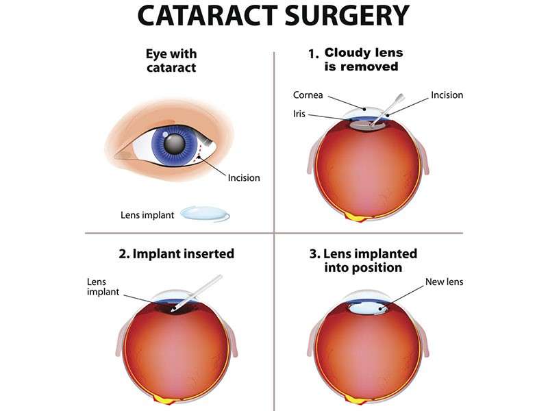
Cataracts develop when proteins in the lens of your eyes clump together causing cloudiness which impairs vision progressively over time. Awareness about this common yet treatable condition is vital as it affects many people as they age making timely intervention essential!
DIABETIC RETINOPATHY
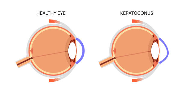
This condition occurs in individuals with diabetes where high blood sugar levels damage retinal blood vessels. It’s important for diabetic patients to monitor their eyes regularly as this disease progresses silently without noticeable symptoms initially.
- Symptoms: Early stages might show no signs; however later stages could present with blurry vision flashers or dark spots in one’s field of view.
- Causes: Persistent high blood glucose primarily contributes by weakening vessel walls leading them either leak fluid causing swelling or bleed into retinal tissue.
- Diagnosis: Comprehensive dilated exams help detect changes in retinal health through specialized imaging technologies like fundus photography.
- Treatment options: Depending upon severity treatment strategies may involve injections into the eye (anti-VEGF therapy) alongside laser surgery aimed at sealing leaks & preventing further damage.
- What to expect: Regular follow-ups are paramount post-treatment monitoring changes & assessing visual function improvements ongoing health management including proper glucose control is essential as well!
The earlier diabetic retinopathy is diagnosed and treated leads towards better outcomes preventing potential blindness down road!
EYE TRAUMA
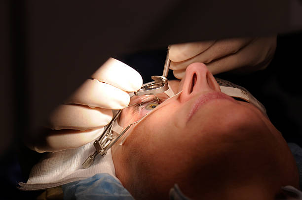
Eyelid or ocular trauma can lead to various injuries affecting vision. Immediate evaluation by a specialist is essential for effective management and minimizing potential long-term effects.
- Symptoms: Symptoms may include visible wounds or cuts around the eye area, bleeding, swelling, impaired vision or inability to open the eyelid completely due to pain.
Seeking immediate medical help following any trauma can significantly influence recovery outcomes.
CORNEAL ULCER
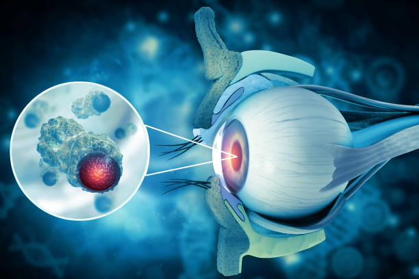
A corneal ulcer is an open sore on the cornea that can result in significant discomfort and vision impairment. Early recognition and appropriate treatment are critical for saving sight.
- Symptoms: Typical signs include severe eye pain, redness, tearing, sensitivity to light, and blurred vision. A whitish spot may also be visible on the cornea.
- Causes: This condition can be caused by infections (bacterial or viral), injury, dry eyes, or improper contact lens use.
- Diagnosis: An ophthalmologist will conduct a comprehensive examination using fluorescein dye which highlights any damage on the cornea's surface under blue light.
- Treatment options: Treatment often includes antibiotic drops for infections or other specific medications based on underlying causes. Severe cases may require surgery.
- What to expect: Recovery time varies; it’s crucial to adhere strictly to prescribed treatments and attend follow-up appointments.
Prompt action against symptoms can prevent serious complications related to corneal ulcers.
ACUTE GLAUCOMA
Acute glaucoma occurs when there is a rapid increase in intraocular pressure, leading to severe pain and potential vision loss. Recognizing its symptoms is vital for timely treatment and prevention of damage.
- Symptoms: Patients often report intense eye pain, headache, nausea, vomiting, blurred vision, and seeing halos around lights.
- Causes: This condition is frequently caused by an obstruction in the drainage system of the eye which can be aggravated by certain medications.
- Diagnosis: An evaluation by an ophthalmologist includes measuring intraocular pressure using tonometry and inspecting the drainage angle with gonioscopy.
- Treatment options: Immediate treatment usually involves medication to lower eye pressure followed by laser therapy or surgery if necessary.
- What to expect: After treatment for acute glaucoma, patients need regular monitoring of their eye pressure and vision to prevent recurrence.
If you suspect you have acute glaucoma symptoms, urgent consultation with an ophthalmologist is imperative.
RETINAL DETACHMENT
Retinal detachment is a serious condition where the retina peels away from its underlying layer of support tissue. This can lead to permanent vision loss if not treated promptly. Understanding the signs and symptoms is crucial for early detection.
- Symptoms: Common indicators include sudden flashes of light, floaters in vision, and a shadow or curtain effect over your sight.
- Causes: Factors that contribute to retinal detachment include aging, previous eye surgery, or trauma to the eye.
- Diagnosis: An ophthalmologist will use a thorough eye examination, including imaging tests like ultrasound or optical coherence tomography (OCT).
- Treatment options: Treatment methods may involve surgical intervention such as scleral buckle or vitrectomy to reattach the retina.
- What to expect: Post-surgery recovery includes rest and limited activity; follow-up visits are essential to monitor healing.
If you experience any of these symptoms, seek medical attention immediately to prevent further complications.