
DR R Tongia
About Me
DR R Tongia
MD
Cardiologist
10 Years Experience
Biography
My Clinic
OPD Hours
| Monday | Morning | 10:00 AM | - | 02:00 PM |
| Evening | 06:00 PM | - | 09:00 PM | |
| Tuesday | Morning | 10:00 AM | - | 02:00 PM |
| Evening | 06:00 PM | - | 09:00 PM | |
| Wednesday | Morning | 10:00 AM | - | 02:00 PM |
| Evening | 06:00 PM | - | 09:00 PM | |
| Thursday | Morning | 10:00 AM | - | 02:00 PM |
| Evening | 06:00 PM | - | 09:00 PM | |
| Friday | Morning | 10:00 AM | - | 02:00 PM |
| Evening | 06:00 PM | - | 09:00 PM | |
| Saturday | Morning | 10:00 AM | - | 02:00 PM |
| Evening | 06:00 PM | - | 09:00 PM | |
| Sunday | Morning | 10:00 AM | - | 02:00 PM |
| Evening | 06:00 PM | - | 09:00 PM |
Treatments
Specialised care and treatments offered
IMPLANTABLE LOOP RECORDER

An implantable loop recorder is a small device placed just beneath the skin that continuously monitors heart rhythms over months or even years. It helps diagnose rare but serious arrhythmias causing unexplained fainting spells or palpitations which are difficult to capture with short-term monitoring techniques.
- PURPOSE AND INDICATIONS: Best suited for patients with infrequent symptoms where traditional external monitors fail to provide conclusive diagnosis.
- TYPICAL PLACEMENT PROCEDURE: Minimally invasive insertion under local anesthesia during an outpatient visit with quick recovery.
- DURATION AND FUNCTIONALITY: Constant ECG recording capability triggers automatic event captures alongside patient-activated recordings transmitted securely for remote analysis.
- BENEFITS AND LIMITATIONS: Provides prolonged monitoring enabling timely diagnosis which guides appropriate management strategies but requires minor surgical implantation.
- TREATMENT DECISIONS FOLLOW-UP: Device data assists cardiologists in determining whether medications adjustment, ablation therapies or pacemaker placement are warranted based on detected arrhythmias.
This advanced diagnostic technology bridges gaps left by conventional assessments offering long-term surveillance essential for complex cardiac symptom evaluation ensuring informed interventions tailored specifically for individual patients’ needs.
CARDIAC STRESS TESTING
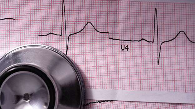
This test evaluates how well the heart performs under physical stress simulated either by exercise or medication when physical activity isn’t possible. Stress testing detects hidden coronary artery disease influencing blood flow during exertion levels increased demand on the heart muscle.
- Syndrome Detection: Identifies angina-related changes when blocked arteries limit oxygen delivery during activity causing chest pain or shortness of breath symptoms triggered only with exertion.
- TYPES OF TESTS: Exercise treadmill test is most common; alternatives include pharmacologic agents mimicking exercise effects if patient unable to perform physical effort.
- METHODS: Continuous ECG monitoring combined with blood pressure measurements assesses cardiac response throughout increasing workload stages.
- NEXT STEPS BASED ON RESULTS: Positive results warrant further imaging like nuclear scans or angiography while negative tests reassure low likelihood of significant blockages.
This functional assessment helps guide individualized treatment plans preventing future cardiac events by uncovering early ischemic changes before they manifest as overt disease signs requiring aggressive intervention.
ECHOCARDIOGRAPHY
Echocardiography uses ultrasound waves to create detailed images of the heart’s structure and function. It is a non-invasive way to assess heart valves, chambers, pumping ability, and detect abnormalities effectively in real time.
- Main Uses: Evaluation of suspected valve diseases, cardiomyopathies, pericardial effusion, or congenital defects causing symptoms like breathlessness or fatigue.
- The Procedure: Performed in-office with an ultrasound probe placed on the chest wall for about 30-45 minutes; painless and radiation-free.
- A Diagnostic Standard: Provides information on ejection fraction and structural integrity essential for treatment decisions including medical therapy or surgery referrals.
Echocardiography remains a cornerstone diagnostic method providing comprehensive data crucial for timely detection and management of cardiovascular conditions without discomfort or risk from invasive testing.
HOLTER MONITORING
This diagnostic tool records cardiac electrical activity continuously over 24-48 hours or longer, identifying intermittent arrhythmias not detected on standard ECGs. It helps in understanding symptoms linked with heart rhythm abnormalities.
- The Purpose: Evaluates palpitations, unexplained dizziness, or fainting episodes potentially due to arrhythmias.
- The Process: A portable device is attached to the chest via electrodes allowing daily activities during monitoring period.
- Data Analysis: Recorded traces reviewed by cardiologists detect frequency, type, and severity of irregular heartbeats.
- Treatment Guidance: Helps tailor medication or decide on further procedures based on findings from continuous rhythm recordings.
This straightforward diagnostic test offers valuable insight into transient cardiac events improving accuracy of diagnosis without hospital admission or invasive methods.
PERCUTANEOUS CORONARY INTERVENTION (PCI)
Blockages in coronary arteries can restrict blood flow leading to chest pain or heart attack. Percutaneous coronary intervention is a minimally invasive method used to open these arteries and restore circulation using balloon angioplasty and stents.
- Symptoms/Causes: Angina, shortness of breath, caused by buildup of plaque narrowing coronary vessels.
- Diagnosis: Stress tests, angiography imaging highlight affected arteries requiring intervention.
- Treatment Approach: A catheter inserted through the groin or wrist delivers balloons and stents to widen arteries.
- Post-Procedure Care: Antiplatelet drugs and lifestyle changes help maintain artery openness and reduce risk of recurrence.
- What to Expect: Relief from symptoms typically within days; hospital stay may be brief depending on complexity.
This intervention provides significant symptomatic improvement while avoiding the need for open-heart surgery in many cases of coronary artery disease.
ATRIAL FIBRILLATION ABLATION
Atrial fibrillation is a common heart rhythm disorder that can cause irregular and often rapid heartbeats. Ablation is a minimally invasive procedure used to correct this by targeting the areas of the heart responsible for abnormal electrical signals.
- Symptoms and Causes: Irregular heartbeat, palpitations, fatigue, and sometimes shortness of breath; caused by erratic electrical impulses in the atria.
- Diagnosis: Electrocardiogram (ECG), Holter monitoring, and sometimes electrophysiological studies identify abnormal rhythms.
- Treatment: Catheter-based ablation uses heat or cold energy to destroy problematic tissue, restoring normal rhythm.
- Management: Post-procedure medications to prevent clots and follow-up monitoring to ensure rhythm stability.
- Expectations: Most patients experience symptom relief within weeks; some may require repeat procedures for full effectiveness.
This technique offers a targeted approach to control atrial fibrillation with a shorter recovery period than open surgery, enhancing quality of life through improved heart function.