
DR Raghupati Sing
About Me
DR Raghupati Sing
MD
Neurologist
10 Years Experience
Biography
My Clinic
OPD Hours
| Monday | Morning | 10:00 AM | - | 02:00 PM |
| Evening | 06:00 PM | - | 09:00 PM | |
| Tuesday | Morning | 10:00 AM | - | 02:00 PM |
| Evening | 06:00 PM | - | 09:00 PM | |
| Wednesday | Morning | 10:00 AM | - | 02:00 PM |
| Evening | 06:00 PM | - | 09:00 PM | |
| Thursday | Morning | 10:00 AM | - | 02:00 PM |
| Evening | 06:00 PM | - | 09:00 PM | |
| Friday | Morning | 10:00 AM | - | 02:00 PM |
| Evening | 06:00 PM | - | 09:00 PM | |
| Saturday | Morning | 10:00 AM | - | 02:00 PM |
| Evening | 06:00 PM | - | 09:00 PM | |
| Sunday | Morning | 10:00 AM | - | 02:00 PM |
| Evening | 06:00 PM | - | 09:00 PM |
Treatments
Specialised care and treatments offered
STROKE RISK ASSESSMENT

An essential evaluation performed to estimate an individual's likelihood of having a stroke by analyzing various controllable and uncontrollable risk factors. This assessment aims at prevention via early intervention strategies.
- Symptoms and causes: Patients with hypertension, atrial fibrillation, diabetes mellitus or family history might need stroke risk assessment despite no current symptoms.
- Diagnosis process: Includes vascular imaging like carotid Doppler ultrasound combined with blood tests assessing cholesterol levels and clotting factors.
- Treatment/management: Lifestyle modifications alongside medications such as antiplatelets or antihypertensives reduce stroke incidence effectively once risk factors identified.
- What to expect: Non-invasive tests conducted over one or two visits provide comprehensive profile guiding personalized preventive measures.
Such evaluations empower patients through proactive health measures reducing long-term disability associated with cerebrovascular events.
MULTIPLE SCLEROSIS WORKUP
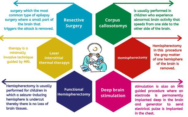
A comprehensive workup for Multiple Sclerosis (MS) comprises clinical evaluation combined with specialized tests aimed at confirming this chronic disease affecting the central nervous system.
- Symptoms and causes: Patients presenting with vision problems, muscle spasms, balance issues or sensory disturbances may undergo an MS workup due to immune-mediated nerve damage suspicion.
- Diagnosis process:MRI scans identify characteristic lesions; lumbar puncture assesses immune markers in cerebrospinal fluid; evoked potentials test nerve pathways' integrity.
- Treatment/management: Early diagnosis allows initiation of disease-modifying therapies minimizing relapses and neurological decline.
- What to expect: Testing spans multiple sessions but yields valuable insights regarding disease activity enabling personalized care planning.
Identifying MS promptly ensures appropriate management strategies aimed at preserving quality of life over time.
NERVE CONDUCTION STUDY (NCS)
The Nerve Conduction Study evaluates how well electrical signals travel through peripheral nerves. This test helps identify nerve damage affecting muscle control and sensation.
- Symptoms and causes: Recommended if experiencing numbness, tingling sensations, muscle weakness, or suspected neuropathies like carpal tunnel syndrome.
- Diagnosis process: Small electrodes stimulate nerves electrically while responses are recorded to measure conduction speed and strength.
- Treatment/management:The outcomes determine nerve injury extent and help monitor diseases such as diabetic neuropathy or nerve compression syndromes.
- What to expect:The procedure usually lasts about an hour with mild discomfort at stimulation sites but no lasting pain.
NCS provides critical information about peripheral nerve function supporting targeted therapeutic interventions for nerve-related conditions.
LUMBAR PUNCTURE (SPINAL TAP)
A lumbar puncture involves extracting cerebrospinal fluid from the lower spine using a thin needle. This diagnostic tool helps detect infections or bleeding around the brain and spinal cord.
- Symptoms and causes: Indicated when patients exhibit signs of meningitis, encephalitis, unexplained headaches, or suspected neurological inflammation.
- Diagnosis process: Performed under sterile conditions with local anesthesia; fluid samples are analyzed for infections or abnormal cells.
- Treatment/management: Results assist in diagnosing infections or autoimmune diseases affecting the nervous system guiding appropriate treatments.
- What to expect: Procedure typically takes 30 minutes; mild post-procedure headache may occur but resolves with hydration and rest.
This procedure remains vital for comprehensive assessment of neurological illnesses through cerebrospinal fluid analysis.
MAGNETIC RESONANCE IMAGING (MRI) OF THE BRAIN
MRI uses strong magnetic fields and radio waves to create detailed images of the brain's internal structures. It is an advanced imaging tool employed to evaluate various neurological disorders.
- Symptoms and causes: Used when symptoms include persistent headaches, weakness, dizziness, or sudden neurological changes indicating possible tumors or lesions.
- Diagnosis process: The patient lies inside a cylindrical scanner; no radiation exposure occurs during this safe procedure.
- Treatment/management: MRI findings may reveal strokes, tumors, infections, or multiple sclerosis aiding in targeted treatment planning.
- What to expect: The scan usually takes 30-60 minutes; patients should avoid metal objects before testing due to strong magnets.
This imaging technique provides critical insights into brain pathology supporting precise diagnosis and therapy guidance.
ELECTROENCEPHALOGRAM (EEG)
An Electroencephalogram, commonly known as EEG, is a diagnostic test that records the electrical activity of the brain. It is a painless and non-invasive procedure widely used to detect abnormal brain wave patterns.
- Symptoms and causes: EEG helps evaluate seizures, unexplained fainting, and epilepsy-related symptoms by monitoring electrical impulses within the brain.
- Diagnosis process: Small electrodes are placed on the scalp to capture brain’s electrical signals while the patient rests or performs specific tasks.
- Treatment/management: Results guide treatment plans for neurological disorders such as epilepsy or sleep disorders.
- What to expect: The test typically lasts 20-40 minutes, requiring minimal preparation; patients can resume normal activities immediately after.
This test is essential for identifying abnormalities in brain function, enabling timely diagnosis and management of neurological conditions.