
Dr Rajendra Chou
About Me
Dr Rajendra Chou
MD
Cardiologist
10 Years Experience
Biography
My Clinic
OPD Hours
| Monday | Morning | 10:00 AM | - | 02:00 PM |
| Evening | 06:00 PM | - | 09:00 PM | |
| Tuesday | Morning | 10:00 AM | - | 02:00 PM |
| Evening | 06:00 PM | - | 09:00 PM | |
| Wednesday | Morning | 10:00 AM | - | 02:00 PM |
| Evening | 06:00 PM | - | 09:00 PM | |
| Thursday | Morning | 10:00 AM | - | 02:00 PM |
| Evening | 06:00 PM | - | 09:00 PM | |
| Friday | Morning | 10:00 AM | - | 02:00 PM |
| Evening | 06:00 PM | - | 09:00 PM | |
| Saturday | Morning | 10:00 AM | - | 02:00 PM |
| Evening | 06:00 PM | - | 09:00 PM | |
| Sunday | Morning | 10:00 AM | - | 02:00 PM |
| Evening | 06:00 PM | - | 09:00 PM |
Treatments
Specialised care and treatments offered
ELECTROPHYSIOLOGICAL STUDY (EPS)

This specialized procedure investigates electrical conduction pathways in the heart to understand abnormal rhythms causing symptoms like fainting or palpitations.
- Symptoms/causes: Indicated for patients with unexplained syncope or complex arrhythmias requiring detailed mapping before ablation therapy.
- Diagnosis: Thin catheters with electrodes are inserted via veins into the heart chambers under fluoroscopic guidance.
- Treatment/management: During EPS, targeted ablation may be performed immediately if arrhythmogenic foci are identified.
- What to expect: This invasive study requires hospital admission with close monitoring; patients may experience temporary discomfort but benefit from definitive treatment options.
EPS provides critical data enabling effective management strategies aimed at restoring normal cardiac rhythm safely.
CARDIAC MRI
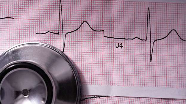
An advanced imaging modality that provides detailed pictures of cardiac anatomy and tissue characteristics without radiation exposure.
- Symptoms/causes:This test assists in evaluating cardiomyopathies, congenital anomalies, myocardial inflammation, and viability before therapies are planned.
- Diagnosis:A powerful magnetic field combined with radio waves generates high-resolution images revealing structural changes and scarring within the myocardium.
- Treatment/management:The insights gained influence decisions related to medical therapy optimization or planning complex surgeries for structural defects。
- What to expect: It is non-invasive and lasts between 30-60 minutes. Some patients require sedation due to enclosed space within the scanner.
Cardiac MRI offers comprehensive information assisting cardiologists in precise diagnosis beyond conventional methods.
CORONARY ANGIOGRAPHY
A coronary angiogram offers precise visualization of coronary arteries to detect blockages that could restrict blood supply to the heart muscle.
- Symptoms/causes: Recommended when there is suspicion of significant coronary artery disease presenting as chest pain or abnormal stress test findings.
- Diagnosis: Involves injecting contrast dye through a catheter inserted into an artery, followed by X-ray imaging of coronary vessels.
- Treatment/management:The findings can lead directly to interventional procedures such as angioplasty or stent placement if needed during the same session.
- What to expect:The procedure is minimally invasive but requires local anesthesia and monitoring; recovery time varies from hours to one day depending on patient condition.
This technique remains the gold standard for detailed assessment of coronary artery blockages enabling targeted treatment strategies.
STRESS TESTING (EXERCISE TREADMILL TEST)
This diagnostic tool assesses how well the heart functions under physical stress by monitoring vital signs during controlled exercise.
- Symptoms/causes: Commonly used in patients with chest discomfort or suspected coronary artery disease.
- Diagnosis: Electrodes attached to the chest record ECG changes while walking on a treadmill with gradually increasing intensity.
- Treatment/management: Results help identify ischemia or arrhythmias triggered by exertion and guide treatment plans such as lifestyle changes or interventions.
- What to expect: The test is supervised closely by medical staff; it typically lasts 10–15 minutes but may be stopped early if symptoms occur.
This non-invasive procedure provides valuable information about cardiovascular performance during physical activity and risk stratification for heart diseases.
24-HOUR HOLTER MONITORING
Continuous heart rhythm monitoring over 24 hours captures intermittent cardiac irregularities that may not appear during a routine ECG.
- Symptoms/causes: Ideal for patients with unexplained palpitations, dizziness, or suspected arrhythmias.
- Diagnosis: A small portable device records electrical activity throughout daily activities to detect abnormal heartbeats.
- Treatment/management: Data assists clinicians in adjusting medications or recommending further interventions based on detected rhythm disturbances.
- What to expect: Patients can maintain usual routines while wearing lightweight leads attached to their chest; results are analyzed after the monitoring period.
This method allows comprehensive evaluation of heart rhythm variability and guides tailored therapeutic decisions accordingly.
ECHOCARDIOGRAPHY
Echocardiography is a widely used imaging technique that helps in evaluating the heart's structure and function through ultrasound waves.
- Symptoms/causes: It is often recommended for patients experiencing symptoms like chest pain, shortness of breath, or arrhythmias to assess cardiac abnormalities.
- Diagnosis: The procedure uses high-frequency sound waves to create live images of the heart valves, chambers, and blood flow patterns.
- Treatment/management: Results help guide treatment plans, including medication adjustments or surgical interventions if structural defects are observed.
- What to expect: Non-invasive and painless, echocardiography usually takes 30–60 minutes and provides detailed insights without radiation exposure.
This test plays a crucial role in diagnosing many cardiovascular conditions by offering real-time visualization of heart activity.