
Dr Rajesh Mishra
About Me
Dr Rajesh Mishra
Ophthalmologist
Biography
My Clinic
OPD Hours
| Tuesday | Morning | 10:00 AM | - | 02:00 PM |
| Evening | 06:00 PM | - | 09:00 PM | |
| Wednesday | Morning | 10:00 AM | - | 02:00 PM |
| Evening | 06:00 PM | - | 09:00 PM | |
| Thursday | Morning | 10:00 AM | - | 02:00 PM |
| Evening | 06:00 PM | - | 09:00 PM | |
| Friday | Morning | 10:00 AM | - | 02:00 PM |
| Evening | 06:00 PM | - | 09:00 PM | |
| Saturday | Morning | 10:00 AM | - | 02:00 PM |
| Evening | 06:00 PM | - | 09:00 PM | |
| Sunday | Morning | 10:00 AM | - | 02:00 PM |
| Evening | 06:00 PM | - | 09:00 PM | |
| Thursday | Morning | 10:00 AM | - | 02:00 PM |
| Evening | 06:00 PM | - | 09:00 PM |
Treatments
Specialised care and treatments offered
OPTICAL COHERENCE TOMOGRAPHY (OCT)
This non-invasive imaging technique provides high-resolution cross-sectional images of the retina and optic nerve head. OCT is pivotal in diagnosing various retinal conditions including macular degeneration and diabetic retinopathy by revealing subtle changes within these structures over time.
- It's particularly useful for detecting issues at an early stage before obvious symptoms arise.
- The test requires minimal preparation; patients simply look into a machine while it captures images without contact.
- Based on results from OCT scans, targeted treatment plans can be developed including injections or other therapies designed specifically around individual needs.
Regular monitoring via OCT offers reassurance and proactive management for patients facing retinal challenges!
CORNEAL TOPOGRAPHY
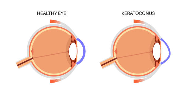
This diagnostic procedure maps the curvature of the cornea's surface using advanced imaging technology. Corneal topography assesses irregularities that could affect vision quality or indicate conditions such as keratoconus or post-surgical changes following procedures like LASIK.
- The primary indications for this test include abnormal vision complaints or preoperative assessments before refractive surgery.
- The procedure is non-invasive; it involves taking multiple measurements across different points on the cornea's surface using specialized equipment.
- The results inform potential management strategies which could involve contact lenses tailored for irregular corneas or further interventions if necessary.
This detailed mapping ensures that patients receive personalized care tailored specifically to their unique corneal shape and refractive errors!
DIABETIC RETINOPATHY SCREENING
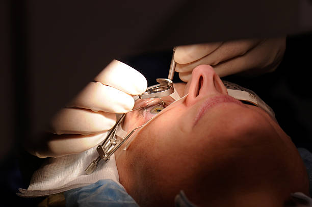
Diabetic retinopathy is an ocular complication of diabetes that affects the blood vessels in the retina. It can lead to severe visual impairment if left untreated. Early detection through screening plays a crucial role in managing this condition effectively.
- This condition often has no early symptoms, making regular screenings essential for diabetic patients.
- A comprehensive dilated eye exam allows an ophthalmologist to assess retinal health and look for signs of damage caused by diabetes.
- If detected early, management options may include lifestyle modifications, laser therapy, or injections that target retinal swelling.<
- Paitents should expect guidance on controlling their diabetes alongside treatment recommendations based on screening outcomes.
Timely screenings can greatly reduce the risk of severe complications associated with diabetic retinopathy!
RETINAL DETACHMENT REPAIR
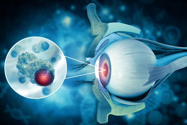
A retinal detachment occurs when the retina separates from its underlying supportive tissue, potentially leading to permanent vision loss if not addressed promptly. Early symptoms may include sudden flashes of light or floaters in the field of vision.
- Main causes include aging, injury, or severe myopia (nearsightedness).
- An ophthalmologist will perform a detailed examination that may involve ultrasound imaging to confirm a diagnosis.
- Treatment options may involve surgical interventions like scleral buckle surgery or vitrectomy to reattach the retina.
- Post-operative care is important for recovery; follow-up visits will ensure proper healing and monitor visual improvements.
Timely intervention is vital for optimal chances of restoring sight after a retinal detachment.
GLAUCOMA MANAGEMENT
Glaucoma refers to a group of eye disorders that cause damage to the optic nerve, often associated with increased intraocular pressure. It is one of the leading causes of blindness worldwide.
- The main symptoms may not be apparent until significant damage has occurred; however, peripheral vision loss can occur over time.
- A thorough eye exam helps with diagnosis, typically involving measuring eye pressure and examining the optic nerve.
- Treatment generally includes medications such as eye drops, laser treatments, or surgical procedures aimed at lowering intraocular pressure.
- Regular check-ups are crucial to monitor progression and adjust treatment as needed.
Persistent management is essential for preserving vision in patients diagnosed with glaucoma.
CATARACT SURGERY
Cataracts are a common eye condition characterized by clouding of the lens, which can lead to blurred vision and difficulty in daily activities. This condition often develops slowly and may not be noticeable initially.
- Symptoms include cloudy or blurry vision, sensitivity to light, and seeing halos around lights.
- Diagnosis is determined through a comprehensive eye examination, including visual acuity tests and a slit-lamp examination.
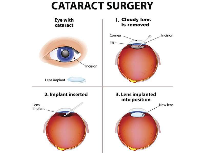
- Treatment options primarily involve surgery, where the cloudy lens is removed and replaced with an artificial intraocular lens.
- Patients can expect improved vision after surgery, although some may require glasses for specific tasks post-operation.
Early detection and surgical intervention can significantly enhance quality of life for those affected by cataracts.