
Dr Rajesh Sharma
About Me
Dr Rajesh Sharma
MS
Orthopaedic Surgeons
10 Years Experience
Biography
My Clinic
OPD Hours
| Monday | Morning | 10:00 AM | - | 02:00 PM |
| Evening | 06:00 PM | - | 09:00 PM | |
| Tuesday | Morning | 10:00 AM | - | 02:00 PM |
| Evening | 06:00 PM | - | 09:00 PM | |
| Wednesday | Morning | 10:00 AM | - | 02:00 PM |
| Evening | 06:00 PM | - | 09:00 PM | |
| Thursday | Morning | 10:00 AM | - | 02:00 PM |
| Evening | 06:00 PM | - | 09:00 PM | |
| Friday | Morning | 10:00 AM | - | 02:00 PM |
| Evening | 06:00 PM | - | 09:00 PM | |
| Saturday | Morning | 10:00 AM | - | 02:00 PM |
| Evening | 06:00 PM | - | 09:00 PM | |
| Sunday | Morning | 10:00 AM | - | 02:00 PM |
| Evening | 06:00 PM | - | 09:00 PM |
Treatments
Specialised care and treatments offered
PEDIATRIC CLUBFOOT CORRECTION
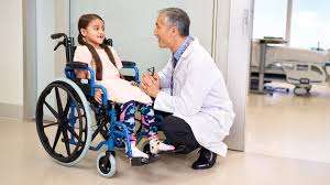
This congenital deformity affects foot positioning resulting in inward twisting that impacts walking ability if untreated. Early intervention aims at correcting foot alignment safely while supporting growth development of affected structures.
- The condition presents as feet turned inward with limited ability to assume normal posture leading parents to seek medical advice shortly after birth.
- A thorough physical exam combined sometimes with X-rays evaluates deformity severity guiding treatment steps which begin promptly after diagnosis for best outcomes.
- Treatment commonly involves serial casting known as Ponseti method that gently stretches tight tendons followed by bracing; surgical release is reserved only when conservative measures fail completely correcting deformity early prevents permanent disability later in life measured follow-ups are essential during treatment course ensuring success over time .
Correcting clubfoot early significantly increases chances children will walk normally minimizing long-term orthopedic issues improving functionality appearance giving families reassurance through structured management
ROTATOR CUFF REPAIR
Tears in the rotator cuff tendons cause shoulder pain and functional limitations. Repair procedures aim to reattach torn tendons back to bone through minimally invasive techniques or open surgery depending on severity.
- Symptoms typically include shoulder pain worsening at night along with weakness during overhead activities due to tendon disruption.
- An ultrasound or MRI scan confirms diagnosis by delineating tear size and tissue quality essential for surgical planning.
- Surgical repair restores continuity using sutures anchored in bone; post-surgery immobilization allows tendon healing before rehabilitation begins gradually restoring motion range strength and stability over months.
- If untreated chronic tears lead to muscle atrophy making repair more challenging emphasizing timely diagnosis is key for optimal results.
A proper treatment plan tailored individually can significantly decrease discomfort while improving shoulder movement through restoration of rotator cuff integrity.
TOTAL HIP REPLACEMENT

This surgical operation replaces a severely damaged hip joint with artificial components to alleviate pain and improve mobility. It is typically reserved for advanced arthritis or significant joint injury unresponsive to conservative treatments.
- The main symptoms addressed include chronic hip pain limiting daily activities along with stiffness reducing range of motion.
- A combination of physical examination findings supported by X-rays assesses the extent of joint deterioration before surgery is considered appropriate.
- The procedure involves removing damaged cartilage and bone then implanting prosthetic parts made from metal, plastic, or ceramic materials designed for durability and compatibility.
- Patients can expect gradual improvement after surgery with physical therapy focusing on strengthening muscles around the new joint for functional recovery.
This intervention profoundly improves quality of life in selected patients suffering from debilitating hip conditions through reliable restoration of joint mechanics.
FRACTURE FIXATION SURGERY

Surgical stabilization of broken bones ensures proper alignment and promotes healing when fractures are complex or displaced. Various fixation devices help maintain bone position as recovery progresses.
- Fractures causing severe pain, deformity, or instability often require surgical intervention for optimal outcomes.
- Imaging studies like X-rays guide the planning process by revealing fracture type, location, and extent of displacement.
- The procedure includes using plates, screws, rods, or nails to hold bone fragments securely together during healing.
- Postoperative care involves immobilization initially followed by physiotherapy to restore strength and function gradually.
This approach is vital for fractures that cannot be effectively managed with casting alone and helps reduce long-term complications such as malunion or nonunion.
SPINAL DECOMPRESSION THERAPY

Designed to relieve pressure on spinal nerves, this therapy addresses conditions such as herniated discs or spinal stenosis. It employs controlled mechanical stretching of the spine to promote healing and reduce nerve irritation.
- The therapy targets symptoms like back pain radiating to arms or legs, numbness, and muscle weakness caused by nerve compression.
- A detailed clinical evaluation complemented by MRI scans helps in identifying candidates who will benefit most from decompression treatment.
- The process involves non-surgical application of traction forces aimed at expanding intervertebral space and enhancing nutrient flow to damaged discs.
- Patients generally undergo multiple sessions over several weeks, experiencing gradual symptom improvement and enhanced mobility.
This method offers a non-invasive option for managing nerve-related spinal issues while avoiding surgery in many cases.
ARTHROSCOPIC KNEE SURGERY

This minimally invasive procedure is used to diagnose and treat joint problems inside the knee. It involves inserting a small camera called an arthroscope into the joint, allowing the surgeon to see and repair damaged tissues without making large incisions.
- Common indications include meniscal tears, ligament injuries, and cartilage damage causing pain or swelling.
- Diagnosis often begins with clinical examination followed by imaging such as MRI; arthroscopy provides direct visualization and confirmation of internal joint structures.
- Treatment during arthroscopy may range from trimming torn cartilage to repairing ligaments or removing loose bodies.
- Recovery usually involves less pain and quicker rehabilitation compared to open surgery, with many patients regaining function within weeks.
This approach offers precise inspection of the knee’s internal structures and effective management of several conditions with minimal tissue disruption.