
Dr Rekha J Paralkar
About Me
Dr Rekha J Paralkar
Cardiologist
Biography
My Clinic
OPD Hours
| Tuesday | Morning | 10:00 AM | - | 02:00 PM |
| Evening | 06:00 PM | - | 09:00 PM | |
| Wednesday | Morning | 10:00 AM | - | 02:00 PM |
| Evening | 06:00 PM | - | 09:00 PM | |
| Thursday | Morning | 10:00 AM | - | 02:00 PM |
| Evening | 06:00 PM | - | 09:00 PM | |
| Friday | Morning | 10:00 AM | - | 02:00 PM |
| Evening | 06:00 PM | - | 09:00 PM | |
| Saturday | Morning | 10:00 AM | - | 02:00 PM |
| Evening | 06:00 PM | - | 09:00 PM | |
| Sunday | Morning | 10:00 AM | - | 02:00 PM |
| Evening | 06:00 PM | - | 09:00 PM | |
| Thursday | Morning | 10:00 AM | - | 02:00 PM |
| Evening | 06:00 PM | - | 09:00 PM |
Treatments
Specialised care and treatments offered
CHEST X-RAY

A chest X-ray is a common imaging test used by cardiologists for assessing cardiac size and shape as well as checking for fluid accumulation around the lungs which may suggest underlying cardiac issues.
- Symptoms & Causes: This exam might be recommended following symptoms like shortness of breath or persistent coughing that could hint at cardiovascular problems.
- Diagnosis: By using low doses of radiation, images are captured quickly allowing visualization of both lungs and heart structures clearly online.
- Treatment Options: While an X-ray itself does not treat conditions directly; results inform subsequent care pathways including possible referrals for echocardiograms if structural anomalies exist.
- What to Expect: Typically taking only minutes without any preparation required; patients stand against an X-ray plate while images are taken from different angles.
The outcome assists healthcare providers greatly by guiding therapeutic strategies based upon observed abnormalities detected in imaging report
( .CARDIAC CATHETERIZATION
This minimally invasive procedure allows doctors to access coronary arteries through blood vessels. It’s primarily used both diagnostically and therapeutically in managing coronary artery disease (CAD).
- Symptoms & Causes: This procedure may be indicated when symptoms like severe angina occur or when non-invasive tests suggest blockages within arteries.
- Diagnosis: During catheterization, a thin tube (catheter) is inserted into an artery and guided toward the heart while X-ray imaging tracks its positioning.
- Treatment Options: Depending on findings during catheterization, patients might undergo angioplasty with stenting right away if blockages are identified.
- What to Expect: Patients generally feel minimal discomfort; recovery varies but many return home within hours after completion of the procedure.
This technique provides critical information regarding blood flow issues while simultaneously offering solutions when necessary.
HOLTER MONITOR
A Holter monitor is a portable device used for continuous monitoring of your heart’s electrical activity over 24-48 hours. It helps diagnose arrhythmias that may not appear during standard testing.
- Symptoms & Causes: Indications for using this monitor include persistent palpitations, dizziness, or unexplained syncope (fainting).
- Diagnosis: Patients wear electrodes connected to the monitor continuously throughout their daily activities for precise data collection about their heart rhythms.
- Treatment Options: Findings guide decisions regarding medications or further investigations needed based on detected abnormalities in heart rhythms.
STRESS TESTING
A stress test assesses how well your heart functions under physical stress. This test can help determine if there are any underlying coronary artery diseases.
- Symptoms & Causes: Symptoms prompting this test often include unexplained chest discomfort or fatigue during exercise.
- Diagnosis: The test usually involves walking on a treadmill while monitoring your heart rate and rhythm through ECG leads attached to your body.
- Treatment Options: Results may indicate the need for further tests or modifications in exercise routines or medications used for management.
- What to Expect: The test duration varies from 15 to 30 minutes; it's safe but might induce temporary fatigue or breathlessness.
Your physician will interpret the results and discuss appropriate next steps based on your specific cardiovascular health status.
ECHOCARDIOGRAM
The echocardiogram is a non-invasive imaging technique that uses sound waves to create detailed images of the heart's structure and function. It plays an essential role in evaluating various heart conditions.
- Symptoms & Causes: Doctors may recommend this test if you exhibit signs of heart disease such as fatigue, swelling in the legs, or irregular heartbeat.
- Diagnosis: A technician places a transducer on your chest that emits sound waves to visualize the heart on a monitor.
- Treatment Options: Based on findings, treatments may include medications, lifestyle changes, or surgical interventions if necessary.
- What to Expect: The procedure typically lasts about 30-60 minutes and is painless, allowing patients to resume normal activities immediately afterward.
This diagnostic imaging offers valuable information for creating comprehensive care plans tailored to individual needs.
ELECTROCARDIOGRAM (ECG)
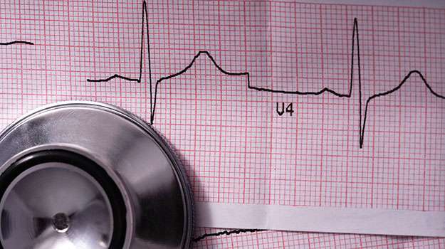
An Electrocardiogram (ECG) is a crucial diagnostic tool used to measure the electrical activity of the heart. It helps identify a range of cardiac issues by recording the timing and strength of electrical signals in the heart.
- Symptoms & Causes: Common reasons for an ECG include chest pain, shortness of breath, palpitations, or fainting spells.
- Diagnosis: An ECG is performed by attaching electrodes to the skin; it captures the heart's activity in real time.
- Treatment Options: While an ECG itself is not a treatment, results can lead to further interventions such as medication adjustments or lifestyle changes.
- What to Expect: The procedure is quick and painless, generally taking 10 minutes or less, with no recovery time needed.
This test provides essential insights that can guide further management and treatment plans for heart conditions.