
Dr Ruchi Jain M
About Me
Dr Ruchi Jain M
DO
Ophthalmologist
10 Years Experience
Biography
My Clinic
OPD Hours
| Monday | Morning | 10:00 AM | - | 02:00 PM |
| Evening | 06:00 PM | - | 09:00 PM | |
| Tuesday | Morning | 10:00 AM | - | 02:00 PM |
| Evening | 06:00 PM | - | 09:00 PM | |
| Wednesday | Morning | 10:00 AM | - | 02:00 PM |
| Evening | 06:00 PM | - | 09:00 PM | |
| Thursday | Morning | 10:00 AM | - | 02:00 PM |
| Evening | 06:00 PM | - | 09:00 PM | |
| Friday | Morning | 10:00 AM | - | 02:00 PM |
| Evening | 06:00 PM | - | 09:00 PM | |
| Saturday | Morning | 10:00 AM | - | 02:00 PM |
| Evening | 06:00 PM | - | 09:00 PM | |
| Sunday | Morning | 10:00 AM | - | 02:00 PM |
| Evening | 06:00 PM | - | 09:00 PM |
Treatments
Specialised care and treatments offered
DIABETIC RETINOPATHY LASER TREATMENT
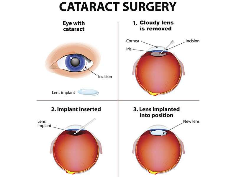
This therapy aims at preventing progression of diabetic retinopathy – a complication of diabetes causing damage to retinal blood vessels – thereby reducing risks of vision impairment or blindness.
- Evolving symptoms may initially be absent but can progress toward blurred vision, floaters, or dark spots as disease advances.
- The diagnosis relies on detailed retinal examination supplemented by fluorescein angiography identifying leaking or abnormal blood vessels requiring treatment.
- Laser photocoagulation selectively seals off leaky vessels and reduces abnormal vessel proliferation responsible for hemorrhages and retinal swelling.
- Multiple sessions could be necessary depending on severity; treatment does not reverse existing damage but prevents worsening significantly.
- Maintaining optimal blood sugar control alongside regular ophthalmic evaluations supports overall management.
Prompt laser intervention alongside systemic diabetic care reduces complications leading to severe visual disability commonly seen without adequate monitoring.
CORNEAL TRANSPLANTATION (KERATOPLASTY)
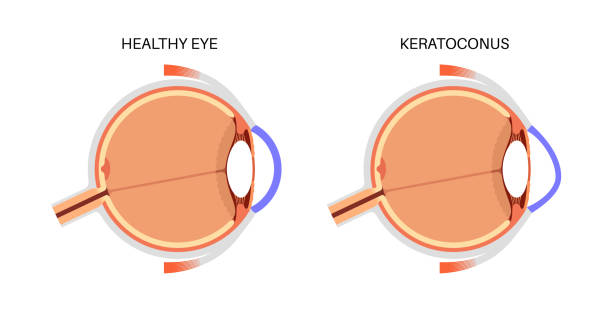
A damaged or diseased cornea leading to impaired vision may require transplantation where diseased tissue is replaced with healthy donor cornea through keratoplasty surgery.
- This condition arises due to scarring, keratoconus, infections, or dystrophies affecting corneal clarity and shape.
- An ophthalmologist evaluates via slit lamp biomicroscopy and pachymetry before surgery planning.
- The procedure entails precise removal of the affected corneal portion and suturing donor tissue in place under sterile conditions in an operating room setting.
- Recovery spans several months involving regular checkups and use of immunosuppressive eye drops to prevent rejection episodes.
Keratoplasty can restore significant visual function but requires patient compliance with postoperative care for successful graft survival long term. Advances in techniques have enhanced safety profiles considerably.
GLAUCOMA FILTERING SURGERY (TRABECULECTOMY)
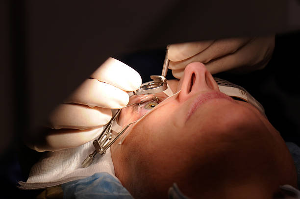
This surgical technique reduces elevated intraocular pressure in glaucoma patients unresponsive to medication, protecting the optic nerve from damage that leads to vision loss.
- Sufferers typically have increased eye pressure detected during routine exams without early noticeable symptoms.
- The procedure creates a new drainage pathway for aqueous humor to lower intraocular pressure effectively.
- Surgery involves forming a small fistula beneath a scleral flap allowing fluid to drain into a filtering bleb under the conjunctiva.
- Postoperative monitoring includes assessments to ensure adequate pressure reduction without complications such as infection or scarring blockage.
This operation can preserve visual function over time when medical management alone fails. Close follow-up care is essential for optimal outcomes.
RETINAL DETACHMENT REPAIR
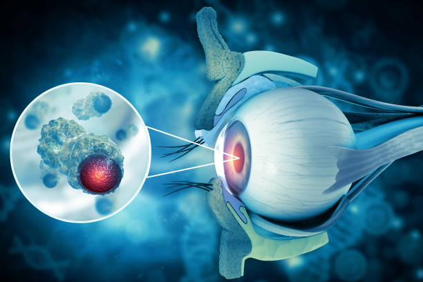
A retinal detachment occurs when the retina separates from its underlying supportive tissue, causing sudden vision loss and requiring urgent treatment to prevent permanent blindness.
- Signs include flashes of light, floaters, shadow in peripheral vision, or curtain-like vision loss.
- The diagnosis is confirmed via dilated fundus examination using specialized imaging techniques like optical coherence tomography (OCT).
- Treatment options range from laser photocoagulation for small tears to surgical interventions such as scleral buckling or vitrectomy for more extensive detachments.
- The goal is to reattach the retina promptly while preserving as much visual function as possible.
Timely intervention improves prognosis dramatically; patients should seek immediate care at any sign of retinal detachment symptoms.
LASIK LASER EYE SURGERY
This innovative procedure corrects refractive errors such as nearsightedness, farsightedness, and astigmatism by reshaping the cornea using laser technology.
- Patients experience symptoms like blurred or distorted vision prior to correction.
- A thorough evaluation including corneal mapping and pupil measurements determines candidacy for LASIK.
- The surgical process involves creating a thin corneal flap followed by laser reshaping of underlying tissues.
- Recovery is generally rapid, with most individuals noticing improved vision within 24 hours.
- Post-surgery precautions include avoiding rubbing eyes and following medication guidelines closely.
LASIK offers a long-term solution to reduce dependency on glasses or contact lenses while maintaining excellent safety standards when performed appropriately.
CATARACT SURGERY
Cataracts cause clouding of the eye's natural lens, leading to blurred vision and potential blindness if untreated. Surgery is often the only effective way to restore clear sight.
- Symptoms include gradual vision loss, glare sensitivity, and faded colors.
- Diagnosis involves a comprehensive eye exam using slit-lamp microscopy and visual acuity tests.
- Treatment consists of removing the cloudy lens and replacing it with an artificial intraocular lens (IOL).
- Procedure is typically performed under local anesthesia and is minimally invasive.
- Postoperative care includes use of prescribed eye drops and avoiding strenuous activity for a short period.
The surgery has a high success rate, significantly improving vision quality for most patients. Early intervention helps prevent complications related to advanced cataracts.