
Dr Sanjeev Garg
About Me
Dr Sanjeev Garg
DO
Ophthalmologist
10 Years Experience
Biography
My Clinic
OPD Hours
| Monday | Morning | 10:00 AM | - | 02:00 PM |
| Evening | 06:00 PM | - | 09:00 PM | |
| Tuesday | Morning | 10:00 AM | - | 02:00 PM |
| Evening | 06:00 PM | - | 09:00 PM | |
| Wednesday | Morning | 10:00 AM | - | 02:00 PM |
| Evening | 06:00 PM | - | 09:00 PM | |
| Thursday | Morning | 10:00 AM | - | 02:00 PM |
| Evening | 06:00 PM | - | 09:00 PM | |
| Friday | Morning | 10:00 AM | - | 02:00 PM |
| Evening | 06:00 PM | - | 09:00 PM | |
| Saturday | Morning | 10:00 AM | - | 02:00 PM |
| Evening | 06:00 PM | - | 09:00 PM | |
| Sunday | Morning | 10:00 AM | - | 02:00 PM |
| Evening | 06:00 PM | - | 09:00 PM |
Treatments
Specialised care and treatments offered
TRAUMATIC HYPHEMA

A hyphema refers to blood accumulation in the front chamber of the eye usually resulting from blunt trauma. Proper assessment and timely management minimize complications like increased intraocular pressure or impaired vision.
- Signs and causes: Visible blood layering in anterior chamber often accompanied by pain following an impact injury to the eye area
- Diagnosis: Slit-lamp examination identifies extent along with monitoring intraocular pressure crucial for detecting secondary glaucoma
- Treatment options: Initial rest with head elevation alongside medicated drops (anti-inflammatory & pressure lowering) controls inflammation & prevents rebleeding
- Patient outlook: Most resolve within weeks without surgery but severe cases require operative evacuation
Careful observation during early post-injury period reduces risk for lasting visual impairment making quick specialized ophthalmic evaluation important following blunt ocular trauma .
ENDOPHTHALMITIS
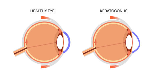
This sight-threatening inflammation inside the eyeball typically arises after surgery or trauma leading to infection of intraocular tissues. Immediate diagnosis and treatment are essential for saving eyesight.
- Pivotal symptoms: Eye pain increasing rapidly postoperatively with redness, reduced vision, swelling around orbit indicate possible endophthalmitis development needing urgent assessment
- Differential diagnosis tools: Clinical examination combined with vitreous sampling helps identify causative organisms guiding targeted antimicrobial therapy
- Treatment modalities: Intravitreal antibiotics injections alongside systemic therapy form mainstay management; sometimes vitrectomy surgery is required in severe infections
CHEMICAL EYE INJURY

An exposure of the eyes to harmful chemicals constitutes an ocular emergency requiring immediate action to limit tissue damage. Rapid irrigation is fundamental first aid before specialist treatment begins.
- Main causes and symptoms: Alkaline or acidic substances contacting the eyes cause redness, pain, tearing, blurred vision and swelling within minutes to hours after exposure.
- Initial evaluation: Eye pH assessment followed by thorough slit-lamp examination determines injury extent including involvement depth beyond surface tissues.
- Treatment strategies: Immediate copious irrigation with saline or water is mandatory; topical antibiotics prevent infection while anti-inflammatory agents reduce scarring risk. Severe cases might need surgical intervention including amniotic membrane grafts.
- Outcome considerations: Prognosis hinges on chemical type and promptness of irrigation; early care decreases risk of blindness significantly.
Avoiding delay in rinsing eyes exposed to chemicals substantially improves chances for functional recovery after these dangerous injuries.
RETINAL DETACHMENT
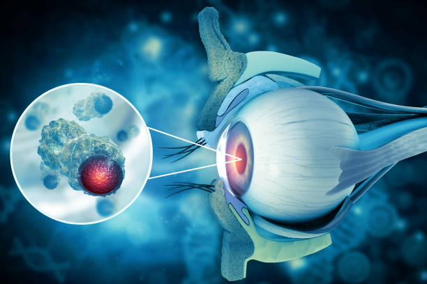
This retinal emergency involves separation of the retina from its underlying layers, threatening permanent vision loss if not treated urgently. Early detection is crucial for successful outcomes.
- Warning signs: New floaters, flashes of light, shadow or curtain over part of vision are common indications of retinal detachment onset.
- Diagnostic methods: Ophthalmologists utilize dilated fundus examination and ocular ultrasound when view is obscured to confirm diagnosis swiftly.
- Treatment interventions: Surgical procedures such as pneumatic retinopexy, scleral buckle, or vitrectomy aim to reattach the retina effectively depending on severity and type of detachment.
- Recovery expectations: Visual improvement varies; prompt repair generally offers better prognosis while delay risks permanent deficits.
If patients notice sudden visual changes described above, urgent ophthalmologic evaluation is vital for preserving sight through appropriate surgical care.
CORNEAL ULCER
A corneal ulcer is an open sore on the cornea often caused by infection or injury. It requires swift medical treatment to avoid complications like scarring or vision loss.
- Symptoms and causes: Painful red eye, tearing, discharge, sensitivity to light, and decreased vision typically indicate an ulcer developing often after trauma or contact lens misuse.
- Diagnosis: Slit-lamp examination with fluorescein dye highlights the ulcer's location and size; cultures may identify infectious agents.
- Treatment approaches: Intensive topical antibiotics or antifungals depending on cause; sometimes steroids are used cautiously under supervision.
- Prognosis: Healing depends on timely therapy; delayed care can lead to corneal scarring affecting vision permanently.
Caring for initial symptoms promptly helps protect eye health and recovery prospects significantly improve with specialized ophthalmic management.
ACUTE ANGLE-CLOSURE GLAUCOMA
This serious eye condition occurs when the drainage angle of the eye becomes suddenly blocked, causing a rapid increase in intraocular pressure. Immediate attention is critical to prevent permanent vision loss.
- Symptoms and causes: Sudden severe eye pain, headache, blurred vision, halos around lights, nausea, and vomiting are common signs due to the abrupt blockage of aqueous humor outflow.
- Diagnosis: An ophthalmologist uses tonometry to measure eye pressure and gonioscopy to inspect the drainage angle for closure.
- Treatment options: Emergency measures include medications to lower eye pressure followed by laser peripheral iridotomy to create a new drainage pathway.
- Management: Prompt treatment usually halts vision damage; if untreated, angle-closure glaucoma can cause irreversible blindness.
Rapid recognition and intervention are essential for preserving sight. Patients experiencing symptoms should seek urgent care without delay.