
About Me
Dr. Shweta Agrawal
DGO DNB OBSGYN
Obstetrics/Gynecology
14 Years Experience
Biography
Dr. Shweta is a highly renowned and skilled Consultant Obstetrician & Gynecologist in Jaipur. She is committed to delivering safe, compassionate, and comprehensive care for women of all ages. With 12+years of clinical experience, she specializes in pregnancy management, high-risk obstetrics, infertility evaluation, gynecological conditions, and advanced minimally invasive surgeries,including Laproscopic ectopic surgery and Laproscopic ovarian cystectomy. Known for her gentle approach and clear communication, Dr. Shweta focuses on understanding each patient’s unique needs and providing treatment that is both evidence-based and personalized. Her dedication to women’s wellness, preventive care, and holistic health makes her a trusted choice for families seeking reliable and empathetic medical guidance.
My Clinic
OPD Hours
Treatments
Specialised care and treatments offered
FIBROID TREATMENT
Uterine fibroids are non-cancerous growths in the uterine wall that can cause various symptoms including heavy periods and pelvic pressure. Treatment options vary based on symptom severity and individual patient goals regarding fertility and overall well-being.
- Common symptoms include excessive menstrual bleeding , discomfort during intercourse, or frequent urination due to pressure on nearby organs.
- They are generally diagnosed through pelvic exams followed by imaging techniques such as ultrasounds.
- Management options range from watchful waiting in asymptomatic cases to medication aimed at shrinking fibroids; surgical treatments like myomectomy could also be considered based on patient needs.
- Many women experience relief once treated successfully; however it's important to discuss personal risks involved with procedures thoroughly beforehand.
Early evaluation helps determine whether management strategies should involve only monitoring or active interventions tailored specifically toward alleviating symptoms effectively while addressing future fertility considerations.
COLPOSCOPY
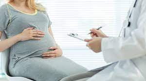
This diagnostic procedure allows further examination of an abnormal Pap smear result using a specialized instrument called a colposcope. It's essential for assessing potential cervical issues accurately before deciding on treatment options.
- It is indicated if there are cellular abnormalities, suspicious lesions noted during routine screenings, or positive HPV tests indicating high risk for cervical cancer.
- The colposcopy provides magnified views of cervical tissues facilitating targeted biopsies when necessary to gather more information about specific areas of concern.
- Treatment might involve further monitoring or direct interventions like cryotherapy if lesions are detected adversely affecting health outcomes.
This examination empowers practitioners with detailed insights regarding cervical health, ensuring timely intervention where required while alleviating patient concerns about abnormal results from earlier tests
ENDOMETRIAL BIOPSY
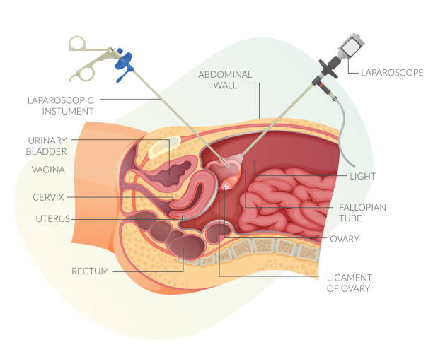
This procedure involves taking a small sample of tissue from the lining of the uterus (endometrium) to assess potential abnormalities. It's critical for diagnosing issues such as unexplained bleeding or evaluating endometrial cancer risk.
- Indications often include heavy menstrual bleeding, irregular cycles, or menopausal bleeding changes.
- The tissue sample is obtained with specialized instruments under local anesthesia and analyzed pathologically for cellular changes or infection signs.
- Treatment following an abnormal biopsy result may involve medication or surgical interventions depending on findings.
- Patiтnts are informed about what to expect during recovery, typically experiencing minor cramping afterward.
An endometrial biopsy provides essential information that guides appropriate treatment plans tailored for individual women's health needs.
PAP SMEAR

A Pap smear is a crucial screening test for cervical cancer and other abnormalities in cervical cells. It plays an important role in early detection, potentially saving lives through preventive measures.
- This test helps identify precancerous changes, which may not show any symptoms initially.
- The process involves collecting cells from the cervix using a brush-like tool, usually during a routine gynecological exam.
- If abnormal results occur, follow-up tests such as HPV testing may be advised to determine further actions needed.
- The Pap smear should be performed regularly as recommended by healthcare professionals based on age and risk factors for optimal health monitoring.
Regular screenings are vital for maintaining reproductive health and preventing serious conditions related to cervical cancer.
HYSTEROSCOPY

This minimally invasive procedure involves inserting a hysteroscope through the cervix into the uterus to diagnose and treat uterine conditions. It allows for direct visualization of the uterine cavity.
- Common indications include abnormal uterine bleeding, fibroids, or polyps.
- The diagnosis is made based on direct inspection of the uterine lining and any anomalies detected.
- Treatment may include removing polyps or fibroids during the same procedure, often leading to immediate improvement of symptoms.
- Patients typically receive local anesthesia and can expect some cramping during the procedure.
Hysteroscopy provides a thorough evaluation of uterine health while minimizing recovery time for patients.
PELVIC ULTRASOUND

A pelvic ultrasound is a non-invasive imaging test that helps visualize the organs within the pelvis, including the uterus and ovaries. It is commonly used to diagnose various conditions affecting women's reproductive health.
- Symptoms such as unexplained pelvic pain, abnormal bleeding, or changes in menstrual cycles may prompt this examination.
- Diagnosis is achieved through high-frequency sound waves that create images of the pelvic organs.
- Treatment options depend on findings; common procedures might include lifestyle modifications or further diagnostic tests.
- Patients can expect to be positioned comfortably as a gel is applied to enhance sound wave transmission during the procedure.
This imaging technique offers valuable insights into reproductive health, aiding in effective treatment decisions.