
Dr Sunil Gupta
About Me
Dr Sunil Gupta
DO
Ophthalmologist
Biography
My Clinic
OPD Hours
| Monday | Morning | 10:00 AM | - | 02:00 PM |
| Evening | 06:00 PM | - | 09:00 PM | |
| Tuesday | Morning | 10:00 AM | - | 02:00 PM |
| Evening | 06:00 PM | - | 09:00 PM | |
| Wednesday | Morning | 10:00 AM | - | 02:00 PM |
| Evening | 06:00 PM | - | 09:00 PM | |
| Thursday | Morning | 10:00 AM | - | 02:00 PM |
| Evening | 06:00 PM | - | 09:00 PM | |
| Friday | Morning | 10:00 AM | - | 02:00 PM |
| Evening | 06:00 PM | - | 09:00 PM | |
| Saturday | Morning | 10:00 AM | - | 02:00 PM |
| Evening | 06:00 PM | - | 09:00 PM | |
| Sunday | Morning | 10:00 AM | - | 02:00 PM |
| Evening | 06:00 PM | - | 09:00 PM |
Treatments
Specialised care and treatments offered
KERATOCONUS
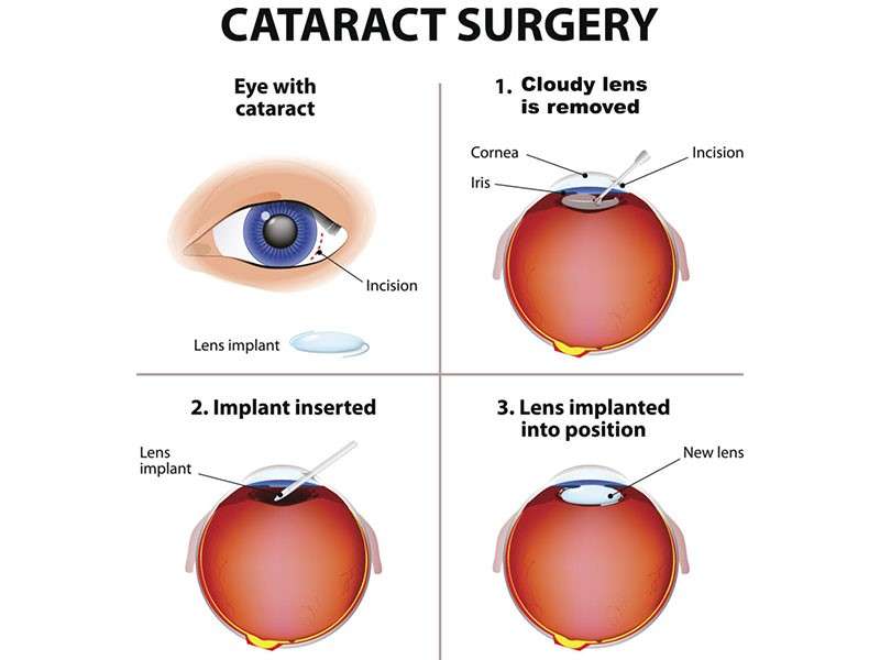
This progressive disorder leads to thinning and bulging of the cornea into a conical shape. Keratoconus primarily affects younger individuals but understanding its implications is vital across all ages if detected early enough.
- Symptoms usually include distorted visions such as blurriness or frequent changes in eyeglass prescriptions arising over time.
- Diagnosis typically involves corneal topography that maps out surface irregularities along with routine examinations focusing on visual acuity measures.
- Management strategies vary depending on severity; mild cases may only require optical corrections while advanced stages could necessitate collagen cross-linking procedures for stabilization or even corneal transplant operations if needed.
Early recognition enables optimal management choices; thus maintaining regular visits encourages proactive care regarding keratoconus – especially beneficial during formative years before complications set too deeply into one’s daily routine!
RETINAL DETACHMENT
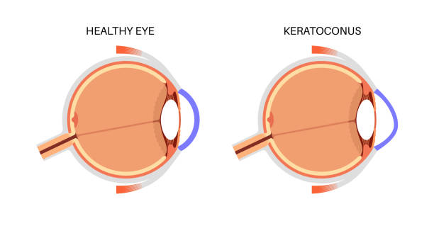
This serious condition occurs when the retina separates from its underlying supportive tissue. Immediate intervention is crucial as it poses a significant risk for permanent blindness if untreated.
- Pain-free symptoms often include sudden onset of floaters or flashes of light and a shadow over your field of vision—these should not be ignored!
- A complete evaluation by an ophthalmologist typically includes visual testing alongside imaging studies like ultrasound to confirm detachment status
Recovery expectations involve follow-up exams to monitor healing progress post-surgery.
AGE-RELATED MACULAR DEGENERATION (AMD)
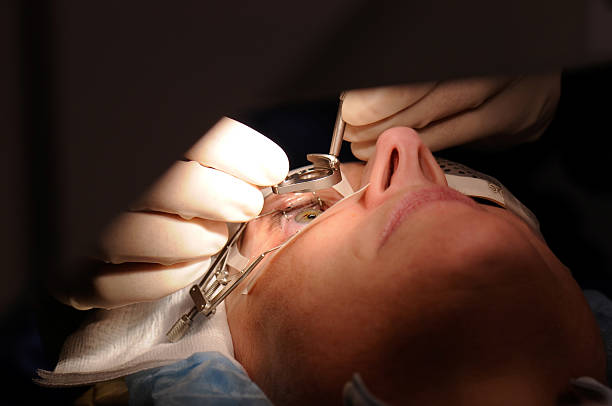
A leading cause of vision loss among older adults, AMD affects the macula—the part of the retina responsible for sharp central vision. Recognizing early signs can aid in effective management.
- Sufferers might notice distortions in straight lines or a gradual decline in central vision clarity as primary symptoms.
- A comprehensive eye exam involving imaging tests helps in early diagnosis by assessing changes within the retina related to AMD.
- While there’s no cure, treatment might include vitamin supplements or injections that slow down disease progression in wet AMD cases.
- Lifestyle modifications such as healthy eating can also play a role in managing this condition effectively
If you experience any changes in your central vision as you age, timely consultations with an ophthalmologist are vital for preserving your eyesight against AMD's effects.
DIABETIC RETINOPATHY
This complication arises from diabetes and affects the blood vessels in the retina. It can lead to serious vision impairment if not addressed early on.
- The condition may present itself with symptoms such as blurred vision, spots or floaters, and difficulty with color perception.
- A thorough examination by an ophthalmologist is essential; tests may include retinal imaging and visual acuity assessments to diagnose its severity.
- Treatment options range from controlling blood sugar levels for prevention to laser therapy or injections for more severe cases where bleeding occurs in the retina.
- Continuous monitoring is crucial for those with diabetes to guard against this potentially sight-threatening complication.
Early detection can significantly improve treatment outcomes for diabetic retinopathy; regular visits are important in safeguarding your vision.
GLAUCOMA
This eye disease is characterized by damage to the optic nerve, often associated with increased intraocular pressure. Glaucoma can lead to irreversible vision loss if not diagnosed and treated promptly.
- Common symptoms include peripheral vision loss, eye pain, headaches, and seeing halos around lights.
- Disease progression is frequently monitored through routine eye examinations that measure eye pressure and assess optic nerve health.
- Treatment strategies may involve prescription eyedrops to lower pressure or surgery in advanced cases to improve drainage of fluid from the eye.
- Regular check-ups are essential for managing this condition effectively and preventing further damage to eyesight.
Your proactive approach in seeking help from an ophthalmologist will be key in preserving your vision from glaucoma's effects.
CATARACTS
Cataracts are a common eye condition that typically develop as people age. This occurs when the lens of the eye becomes cloudy, leading to blurred vision and other visual disturbances.
- Symptoms may include gradual vision loss, double vision, glare from lights, and difficulty seeing at night.
- Cataracts are often diagnosed through a comprehensive eye exam, which includes vision tests and the evaluation of the lens.
- Treatment options typically involve surgical removal of the cloudy lens, which is then replaced with an artificial intraocular lens.
- Patients can expect improved vision after surgery, but it may take time for complete recovery and adjustment to the new lens.
If you experience symptoms of cataracts, consulting an ophthalmologist can help determine the best course of action for your eye health.