
DR V V Agrawal
About Me
DR V V Agrawal
MD
Cardiologist
10 Years Experience
Biography
My Clinic
OPD Hours
| Monday | Morning | 10:00 AM | - | 02:00 PM |
| Evening | 06:00 PM | - | 09:00 PM | |
| Tuesday | Morning | 10:00 AM | - | 02:00 PM |
| Evening | 06:00 PM | - | 09:00 PM | |
| Wednesday | Morning | 10:00 AM | - | 02:00 PM |
| Evening | 06:00 PM | - | 09:00 PM | |
| Thursday | Morning | 10:00 AM | - | 02:00 PM |
| Evening | 06:00 PM | - | 09:00 PM | |
| Friday | Morning | 10:00 AM | - | 02:00 PM |
| Evening | 06:00 PM | - | 09:00 PM | |
| Saturday | Morning | 10:00 AM | - | 02:00 PM |
| Evening | 06:00 PM | - | 09:00 PM | |
| Sunday | Morning | 10:00 AM | - | 02:00 PM |
| Evening | 06:00 PM | - | 09:00 PM |
Treatments
Specialised care and treatments offered
BLOOD PRESSURE MONITORING (AMBULATORY)

Pain-free ambulatory blood pressure monitoring tracks blood pressure continuously over 24 hours outside a clinical environment. It helps diagnose hypertension more accurately than single readings at a clinic visit alone.
- Symptoms/Causes: Used when office measurements are inconsistent with symptoms like headaches or fatigue suggesting high blood pressure effects.
- Diagnosis Process: A cuff attached to a portable monitor inflates periodically throughout day and night recording pressures during various activities including sleep.
- Treatment/Management: Provides comprehensive data allowing optimization of antihypertensive drugs tailored to daily fluctuations.
- What To Expect: Patients can go about routine tasks but should keep arm still during measurements; results inform ongoing cardiovascular risk management.
This method ensures better control of hypertension reducing long-term risks of stroke and heart attack through more individualized care plans.
CORONARY ANGIOGRAPHY
This diagnostic procedure visualizes blood flow through coronary arteries using contrast dye and X-ray imaging. It identifies blockages causing angina or myocardial infarction risk.
- Symptoms/causes: Indicated when symptoms like persistent chest pain suggest significant coronary artery disease unconfirmed by non-invasive tests.
- Diagnosis process: A catheter inserted via an artery delivers contrast dye into coronary vessels while fluoroscopy produces real-time imagery of arterial patency and obstructions.
- Treatment/Management:The findings guide interventional treatments such as angioplasty with stent placement or coronary bypass surgery recommendations.
HOLTER MONITORING
A Holter monitor is a portable device worn for 24-48 hours to continuously record the heart’s electrical activity during normal daily routines. It detects intermittent arrhythmias that a single ECG may miss.
- Symptoms/causes: Recommended for patients experiencing unexplained palpitations, syncope episodes, or irregular heartbeat sensations occurring sporadically.
- Diagnosis process: Electrodes connected to a small recording device capture continuous ECG data while patients continue their usual activities.
- Treatment/Management: Data analysis helps tailor antiarrhythmic therapy or identify needs for pacemaker implementation if necessary.
- What to expect: Patients should maintain normal activities but avoid bathing; wearing it can cause minor skin irritation from electrodes in some cases.
This convenient monitoring technique allows detection of transient cardiac conduction problems that might otherwise be missed during brief clinical assessments.
STRESS TESTING
A stress test evaluates how well the heart performs under physical exertion. This test detects coronary artery disease and exercise-induced arrhythmias that may not appear when at rest.
- Symptoms/causes: Patients with chest pain during activity or risk factors for heart disease are typical candidates.
- Diagnosis process: Monitors ECG changes while the patient walks on a treadmill or pedals a stationary bike; medications can be used if exercise isn’t possible.
- Treatment/Management: Helps determine the need for lifestyle changes, medications, or further invasive procedures like angiography.
- What to expect: The procedure involves progressively increasing exercise intensity under medical supervision; it's generally safe with continuous monitoring of vital signs.
This dynamic assessment provides critical information about myocardial blood flow and cardiac response to exertion, assisting in risk stratification and treatment planning.
ECHOCARDIOGRAPHY
Echocardiography uses ultrasound waves to create detailed images of the heart’s chambers, valves, and surrounding structures. It provides both anatomical and functional information.
- Symptoms/causes: It is often advised for patients with murmurs, unexplained breathlessness, or suspected valve disease.
- Diagnosis process: A transducer placed on the chest sends sound waves that echo off cardiac tissues to form live images on a monitor.
- Treatment/Management: Identifies issues such as valve dysfunction or cardiomyopathy, aiding decisions regarding medical or surgical interventions.
- What to expect: The test is safe, non-invasive, usually lasts 30-45 minutes, and requires no special preparation.
This imaging technique is invaluable in assessing cardiac anatomy and guiding management strategies effectively.
ELECTROCARDIOGRAM (ECG)
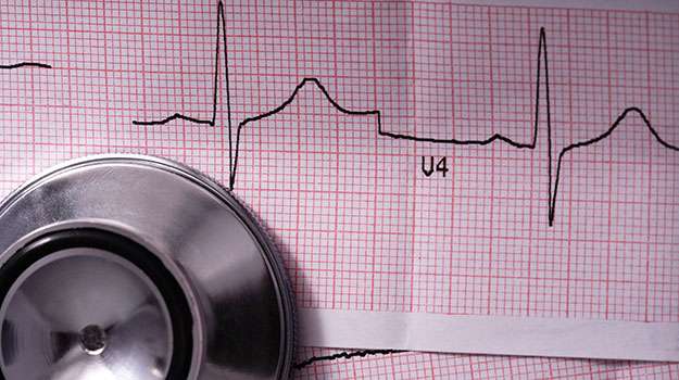
An Electrocardiogram (ECG) is a fundamental diagnostic test used in cardiology to assess the heart's electrical activity. It helps detect abnormalities in heart rhythm and structure.
- Symptoms/causes: It is typically performed when patients experience chest pain, palpitations, dizziness, or unexplained shortness of breath.
- Diagnosis process: Electrodes placed on the skin record the electrical impulses generated by the heart, producing wave patterns on paper or a screen.
- Treatment/Management: Results guide treatment plans, such as medication adjustments for arrhythmias or further testing for ischemic heart disease.
- What to expect: The procedure is painless, takes about 5-10 minutes, and requires minimal preparation.
This non-invasive test provides quick insights into cardiac function and is an essential first step in evaluating many heart conditions.