
Dr. Saurabh Heart Clinic
About Me
Dr. Saurabh Heart Clinic
MD
Cardiologist
10 Years Experience
Biography
My Clinic
OPD Hours
| Monday | Morning | 10:00 AM | - | 02:00 PM |
| Evening | 06:00 PM | - | 09:00 PM | |
| Tuesday | Morning | 10:00 AM | - | 02:00 PM |
| Evening | 06:00 PM | - | 09:00 PM | |
| Wednesday | Morning | 10:00 AM | - | 02:00 PM |
| Evening | 06:00 PM | - | 09:00 PM | |
| Thursday | Morning | 10:00 AM | - | 02:00 PM |
| Evening | 06:00 PM | - | 09:00 PM | |
| Friday | Morning | 10:00 AM | - | 02:00 PM |
| Evening | 06:00 PM | - | 09:00 PM | |
| Saturday | Morning | 10:00 AM | - | 02:00 PM |
| Evening | 06:00 PM | - | 09:00 PM | |
| Sunday | Morning | 10:00 AM | - | 02:00 PM |
| Evening | 06:00 PM | - | 09:00 PM |
Treatments
Specialised care and treatments offered
TRANSESOPHAGEAL ECHOCARDIOGRAM (TEE)

A transesophageal echocardiogram (TEE) provides more detailed images of the heart compared with standard echocardiograms by passing an ultrasound probe down into the esophagus. It's particularly useful when clearer images are needed due to patient anatomy or other challenges with traditional methods
- Symptoms may include suspicion of clots forming within chambers or detection issues with valves seen via regular echocardiograms
- A light sedation ensures comfort while placing this probe carefully into position before capturing high-resolution images within cardiology settings offering maximum diagnostic precision
- It focuses mainly on left atrial appendage assessments aiding greatly concerning pre-operative planning amongst patients needing surgery due excessive clot risks previously presented
- Treatment plans grow tailored according ultimately depending identified concerns noted throughout evaluation period following sedated procedures regularly conducted safely
- TEE usually requires post-procedure observations allowing recovery; however standards protocols assure rapid transitions back towards normality soon thereafter allowing practitioners gain actionable insights rapidly enhancing therapy pathways overall
Opting for TEE guarantees comprehensive evaluations ensuring professional expertise maximizes evolving cardiac care strategies flawlessly moving forward effectively
CARDIAC CT ANGIOGRAPHY
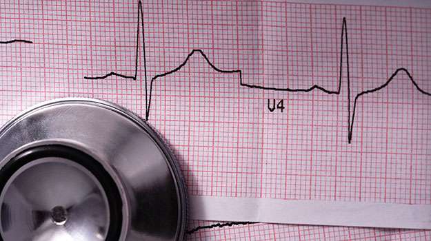
Cardiac CT angiography utilizes advanced imaging technology to visualize blood vessels supplying blood to the heart. This non-invasive technique is effective in detecting blockages that could lead to serious cardiovascular problems.
- Indications for this exam often stem from symptoms like significant chest pain or abnormal stress tests indicating potential ischemia risk.
- The patient will undergo scanning after receiving an intravenous contrast dye which enhances visualization during imaging
- Diagnosis hinges upon assessing any narrowed arteries found within these detailed images captured during the procedure
- Management strategies might range from simple observation through higher-level interventions such as stenting if significant obstructions are revealed
- Patients typically receive thorough instructions prior regarding preparation requirements ensuring optimal results
Having a Cardiac CT angiogram can lead directly towards timely interventions thereby improving long-term outcomes considerably
STRESS TESTING
A stress test evaluates how well your heart performs under physical pressure. This assessment helps identify potential coronary artery disease or other cardiac problems that may not be evident at rest.
- Common symptoms leading to this assessment might include chest discomfort during exertion or unusual fatigue levels while performing routine activities.
- The procedure involves exercising on a treadmill while monitoring your heart rate and rhythm through EKG leads attached to your body.
- If exercise isn't possible, medications can simulate stress on the heart while being monitored closely by healthcare staff.
- Results will guide treatment decisions—ranging from lifestyle modifications to potential referrals for more invasive procedures if necessary.
- This diagnostic method typically lasts around 30 minutes and is performed in a controlled clinical environment ensuring patient safety at all times.
A stress test is an essential component in evaluating overall cardiovascular fitness and health risks effectively!
HOLTER MONITOR
A Holter monitor is a portable device that continuously records the heart's rhythm for 24 to 48 hours. It is crucial for diagnosing intermittent cardiac issues that may not appear during standard tests.
- Symptoms prompting this test include palpitations, dizziness, or fainting spells without clear explanation.
- The patient wears electrodes connected to a small recording device throughout their normal daily activities.
- The data collected aids in identifying arrhythmias or conduction disorders that could impact heart health.
- Treatment options will depend on findings; some patients may require medication adjustment or lifestyle changes based on results.
- This test generally does not disrupt daily activities and allows for a comprehensive overview of cardiac function over time.
ECHOCARDIOGRAM
An echocardiogram uses ultrasound waves to create images of the heart's structures and functions. This non-invasive test helps in diagnosing various cardiac conditions.
- Common indications include unexplained shortness of breath, fatigue, or symptoms suggesting heart valve problems.
- The test is performed by placing a transducer on the chest to capture real-time images of the heart.
- It provides critical information about heart muscle thickness, chamber size, and overall function.
- Treatment approaches may vary depending on the results but can include medication or further testing if necessary.
- This safe procedure usually takes about 30-60 minutes with no downtime required afterward.
Echocardiograms are essential tools in cardiology for monitoring heart health effectively.
CORONARY ANGIOGRAPHY
Coronary angiography is a diagnostic procedure used to visualize the arteries of the heart. This technique allows doctors to assess the presence and extent of coronary artery disease.
- Symptoms may include chest pain, shortness of breath, or other signs of heart issues.
- The procedure is typically carried out under local anesthesia and involves inserting a catheter into an artery.
- Diagnosis is confirmed by observing the flow of contrast dye through the coronary arteries using X-ray imaging.
- Treatment options may include lifestyle changes, medication, or surgical intervention based on the findings.
- Patients can expect to be monitored closely before, during, and after the procedure for their safety.
This test is vital for assessing heart health and determining appropriate treatment strategies.