
drmamta jain
About Me
drmamta jain
DO
Ophthalmologist
10 Years Experience
Biography
My Clinic
OPD Hours
| Monday | Morning | 10:00 AM | - | 02:00 PM |
| Evening | 06:00 PM | - | 09:00 PM | |
| Tuesday | Morning | 10:00 AM | - | 02:00 PM |
| Evening | 06:00 PM | - | 09:00 PM | |
| Wednesday | Morning | 10:00 AM | - | 02:00 PM |
| Evening | 06:00 PM | - | 09:00 PM | |
| Thursday | Morning | 10:00 AM | - | 02:00 PM |
| Evening | 06:00 PM | - | 09:00 PM | |
| Friday | Morning | 10:00 AM | - | 02:00 PM |
| Evening | 06:00 PM | - | 09:00 PM | |
| Saturday | Morning | 10:00 AM | - | 02:00 PM |
| Evening | 06:00 PM | - | 09:00 PM | |
| Sunday | Morning | 10:00 AM | - | 02:00 PM |
| Evening | 06:00 PM | - | 09:00 PM |
Treatments
Specialised care and treatments offered
EMERGENCY EYE CARE
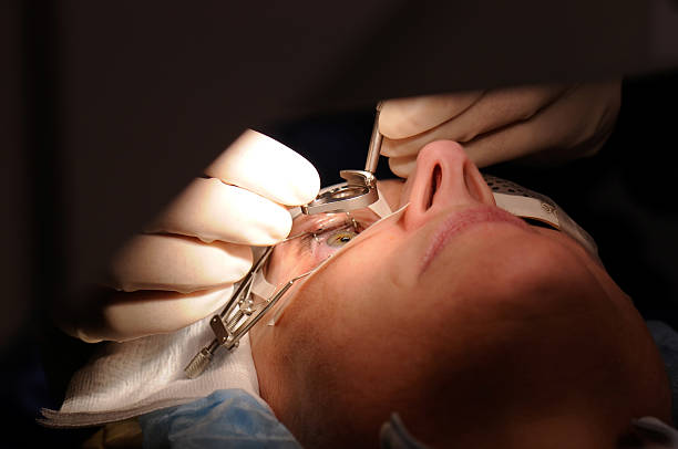
Eye emergencies can arise unexpectedly from injuries, chemical exposures, or sudden changes in vision. Immediate medical attention is paramount for optimal outcomes in such situations!
- Symptoms demanding urgent evaluation include severe pain, sudden loss of vision, or visible foreign objects in the eye.
- Diagnosis will entail a thorough examination including imaging studies if needed depending on injury severity.
- Initial management may vary widely—from flushing chemicals out of eyes to suturing lacerations—based on specific circumstances encountered during evaluation.
- Follow-up care isn't just important; it helps ensure complete recovery while monitoring for any potential long-term effects.
Being prepared with knowledge about potential emergencies enables quicker reactions which could save your sight!
KERATOCONUS MANAGEMENT
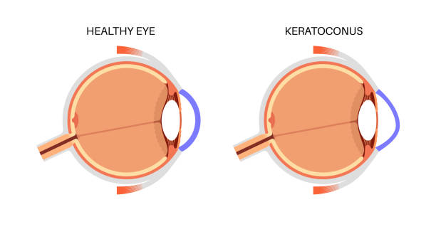
Keratoconus is a progressive eye disorder where the cornea thins and bulges into a cone shape causing distorted vision. Awareness about early signs can help slow its progression with timely interventions.
- The initial symptoms often include blurry eyesight that worsens over time alongside increased sensitivity to light and glare issues at night.
- An ophthalmologist diagnoses keratoconus through corneal topography which maps out corneal curvature and thickness accurately.
- Treatment options generally begin with special contact lenses but may escalate to corneal cross-linking procedures or even transplantation if more severe stages occur.
- Regular monitoring is crucial for adapting treatment plans based on disease progression.
Prompt recognition allows for tailored management strategies that can stabilize visual function significantly.
DIABETIC RETINOPATHY TREATMENT
This complication of diabetes affects blood vessels in the retina and can lead to blindness without appropriate care. Early detection makes a significant difference in treatment outcomes.
- Symptoms may range from no noticeable changes in vision to blurred or fluctuating eyesight as well as dark spots in one's visual field.
- A comprehensive diabetic exam typically reveals any retinal changes indicative of diabetic retinopathy through fundoscopy or fluorescein angiography.
- Treatment strategies include laser therapy, anti-VEGF injections, or vitrectomy based on disease progression.
- Lifestyle modifications such as better blood sugar control can also play a vital role in managing this condition effectively.
Your proactive engagement with regular screening can help maintain your precious sight despite diabetes complications.
RETINAL DETACHMENT REPAIR
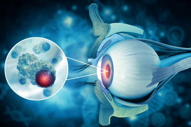
A retinal detachment is a serious condition that occurs when the retina separates from its underlying supportive tissue. Prompt treatment is essential to prevent permanent loss of vision.
- Symptoms often include sudden flashes of light, floaters, or a shadow moving across your field of vision.
- An ophthalmologist will diagnose this condition through a dilated eye exam and imaging tests like optical coherence tomography (OCT).
- Treatment options may include laser surgery, cryopexy (freezing), or vitrectomy depending on the severity of detachment.
- Recovery requires careful adherence to post-operative instructions to ensure optimal results.
Understanding this condition allows for timely intervention which can safeguard your sight from irreversible damage.
GLAUCOMA MANAGEMENT
Glaucoma is often dubbed the 'silent thief of sight' due to its gradual onset and lack of early symptoms. It occurs when increased pressure in the eye damages the optic nerve, potentially leading to permanent vision loss.
- Common causes include elevated intraocular pressure, family history, and certain medical conditions like diabetes.
- A comprehensive eye examination with tonometry helps diagnose glaucoma by measuring intraocular pressure.
- Treatment options may involve prescription eye drops, oral medications, or surgical procedures aimed at lowering eye pressure.
- Routine monitoring is essential for managing glaucoma effectively and preventing further damage.
Early detection is crucial; regular check-ups can significantly contribute to preserving your vision.
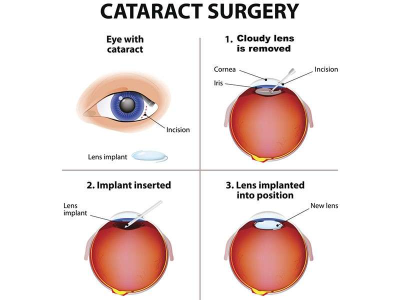
CATARACT SURGERY
Cataracts are a common condition that affects the clarity of the eye's lens, leading to blurred vision. They are often age-related but can also be caused by trauma or certain medications.
- Typical symptoms include cloudy or blurry vision, difficulty seeing at night, and sensitivity to light.
- Cataracts are diagnosed during a comprehensive eye exam where the doctor assesses vision clarity and examines the lens.
- Treatment primarily involves surgical removal of the cloudy lens, with an artificial intraocular lens implanted to restore vision.
- Post-operative care may include eye drops and follow-up visits to ensure proper healing.
- Most patients experience a significant improvement in their eyesight following surgery.
Timely intervention can help regain clear vision and enhance quality of life.