
DrSanjay KuMar S
About Me
DrSanjay KuMar S
DO
Ophthalmologist
10 Years Experience
Biography
My Clinic
OPD Hours
| Monday | Morning | 10:00 AM | - | 02:00 PM |
| Evening | 06:00 PM | - | 09:00 PM | |
| Tuesday | Morning | 10:00 AM | - | 02:00 PM |
| Evening | 06:00 PM | - | 09:00 PM | |
| Wednesday | Morning | 10:00 AM | - | 02:00 PM |
| Evening | 06:00 PM | - | 09:00 PM | |
| Thursday | Morning | 10:00 AM | - | 02:00 PM |
| Evening | 06:00 PM | - | 09:00 PM | |
| Friday | Morning | 10:00 AM | - | 02:00 PM |
| Evening | 06:00 PM | - | 09:00 PM | |
| Saturday | Morning | 10:00 AM | - | 02:00 PM |
| Evening | 06:00 PM | - | 09:00 PM | |
| Sunday | Morning | 10:00 AM | - | 02:00 PM |
| Evening | 06:00 PM | - | 09:00 PM |
Treatments
Specialised care and treatments offered
CATARACTS
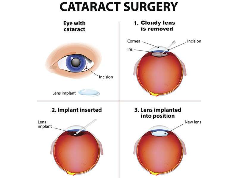
A cataract forms when proteins in the lens clump together causing cloudiness which affects clear vision over time. This common condition usually develops gradually but can significantly impair daily activities if left untreated
.- .
OCULAR TRAUMA
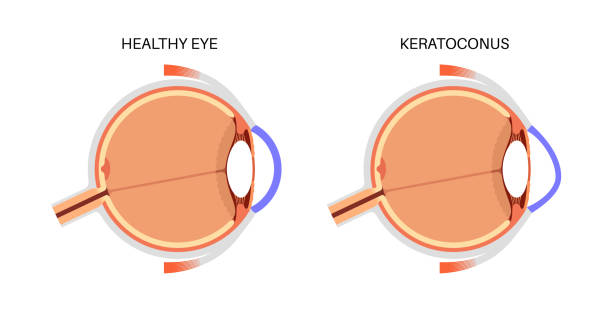
An injury affecting any part of the eye can lead to ocular trauma which may result in severe complications if not addressed immediately. Awareness about potential injuries during activities can help prevent such incidents).
ACUTE ANGLE-CLOSURE GLAUCOMA
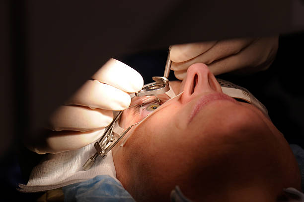
This urgent condition arises when there is a sudden increase in intraocular pressure due to blockage of fluid drainage from the eye. It requires immediate intervention to prevent permanent vision loss associated with it.
- Symptoms: Sudden onset of severe headache, eye pain, nausea, vomiting, and blurred vision are common presentations.
- Causes: Typically occurs in people with narrow anterior chamber angles that predispose them to blockages.
- Diagnosis: An ophthalmologist evaluates symptoms along with measuring intraocular pressure during an emergency visit.
- Treatment Options: Urgent treatments include medication for lowering pressure followed by laser or surgical intervention quickly thereafter.
- What to Expect: Quick relief from pain after treatment but close follow-up care will be important for managing future risks.
Prompt action upon noticing symptoms is vital for preserving eyesight when it comes to acute angle-closure glaucoma.
DIABETIC RETINOPATHY
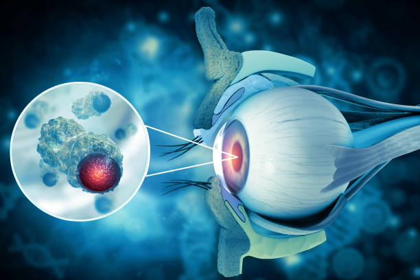
This complication of diabetes affects the blood vessels in the retina and can result in vision impairment over time. Early detection is critical in preventing severe outcomes associated with this condition.
- Symptoms: Blurred vision, floaters, or difficulty seeing at night may indicate diabetic retinopathy's onset.
- Causes: Prolonged high blood sugar levels damage retinal blood vessels leading to swelling and leakage of fluids into the retina.
- Diagnosis: A dilated eye exam allows for thorough observation of changes in the retina that are characteristic of this condition.
- Treatment Options: Options include laser treatment to seal leaking blood vessels or injections of medications that reduce swelling in the retina.
- What to Expect:Please follow up regularly with your ophthalmologist for monitoring and adjustment of treatment as needed based on diabetes management efforts.
GLAUCOMA
This progressive eye disease is characterized by increased intraocular pressure that can damage the optic nerve. Glaucoma can lead to irreversible vision loss if not detected early and managed appropriately.
- Symptoms: Many patients may not notice symptoms until significant damage has occurred; however, peripheral vision loss is common.
- Causes: It often arises due to fluid buildup in the eye but may also be hereditary or linked with other medical conditions.
- Diagnosis: Comprehensive eye exams measure intraocular pressure and assess the health of the optic nerve using visual field tests and OCT scans.
- Treatment Options: Management may involve prescription eye drops, oral medications, laser therapy, or surgical procedures aimed at lowering eye pressure.
- What to Expect: Regular monitoring will be necessary as glaucoma is a lifelong condition requiring ongoing management to preserve vision.
If diagnosed early, the progression of glaucoma can often be effectively managed with timely treatments.
RETINAL DETACHMENT
Retinal detachment is a serious condition where the retina separates from its underlying supportive tissue. This can lead to permanent vision loss if not treated promptly. It often occurs without any warning, making awareness of its symptoms crucial.
- Symptoms: Patients may experience sudden flashes of light, floaters, or a shadow over their vision.
- Causes: Common causes include extreme nearsightedness, previous eye surgery, or trauma.
- Diagnosis: Eye examinations using a slit-lamp and retinal imaging techniques help in identifying the detachment.
- Treatment Options: Surgical interventions such as pneumatic retinopexy, scleral buckling, or vitrectomy are commonly used to reattach the retina.
- What to Expect: Post-surgery recovery includes follow-up visits and some restrictions on activities to ensure proper healing.
If you notice any symptoms indicative of retinal detachment, it's imperative to seek immediate medical attention.