
Dr Alok Mathur -
About Me
Dr Alok Mathur -
MD
Cardiologist
10 Years Experience
Biography
My Clinic
OPD Hours
| Monday | Morning | 10:00 AM | - | 02:00 PM |
| Evening | 06:00 PM | - | 09:00 PM | |
| Tuesday | Morning | 10:00 AM | - | 02:00 PM |
| Evening | 06:00 PM | - | 09:00 PM | |
| Wednesday | Morning | 10:00 AM | - | 02:00 PM |
| Evening | 06:00 PM | - | 09:00 PM | |
| Thursday | Morning | 10:00 AM | - | 02:00 PM |
| Evening | 06:00 PM | - | 09:00 PM | |
| Friday | Morning | 10:00 AM | - | 02:00 PM |
| Evening | 06:00 PM | - | 09:00 PM | |
| Saturday | Morning | 10:00 AM | - | 02:00 PM |
| Evening | 06:00 PM | - | 09:00 PM | |
| Sunday | Morning | 10:00 AM | - | 02:00 PM |
| Evening | 06:00 PM | - | 09:00 PM |
Treatments
Specialised care and treatments offered
VASCULAR DISEASES
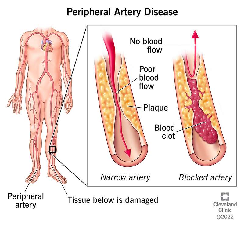
Vascular Diseases encompass a range of conditions affecting the blood vessels, including peripheral artery disease (PAD), carotid artery disease, and venous disorders. Effective management aims to alleviate symptoms, prevent complications, and improve quality of life.
🩺 Lifestyle Modifications
Adopting a heart-healthy lifestyle is foundational in managing vascular diseases:
- Smoking Cessation: Quitting smoking is crucial, as tobacco use accelerates atherosclerosis and increases the risk of cardiovascular events .prathimahospitals.com
- Dietary Changes: Emphasizing fruits, vegetables, whole grains, and lean proteins while reducing saturated fats helps manage cholesterol levels and blood pressure.
- Regular Exercise: Engaging in physical activity improves circulation and overall vascular health.
- Weight Management: Maintaining a healthy weight reduces strain on the cardiovascular system.
💊 Medications
Pharmacological interventions are tailored to individual needs:
- Antiplatelet Agents: Medications like aspirin or clopidogrel help prevent blood clots, reducing the risk of heart attacks and strokes .
- Statins: These drugs lower cholesterol levels, stabilizing plaques and preventing vascular events.
- Antihypertensives: Controlling blood pressure with medications such as ACE inhibitors or beta-blockers is essential in managing vascular health.
- Diabetes Management: Maintaining blood sugar levels within target ranges reduces the risk of complications in diabetic patients with vascular issues.
🥠Surgical and Interventional Procedures
In advanced cases, surgical interventions may be necessary:
- Angioplasty and Stenting: A balloon is used to open narrowed arteries, and a stent may be placed to keep the artery open .
- Atherectomy: Plaque is removed from the arteries to restore blood flow.
- Bypass Surgery: A graft is used to reroute blood around blocked arteries.
- Endarterectomy: Surgical removal of fatty deposits from artery walls.
- Thrombolytic Therapy: Clot-dissolving drugs are administered to treat acute blockages
🩹 Wound Care and Compression Therapy
For conditions like venous ulcers or PAD-related wounds:
- Wound Care: Proper cleaning and dressing of ulcers promote healing.
- Compression Therapy: Using compression stockings helps reduce swelling and improve blood flow in venous diseases .
Conclusion
Managing vascular diseases requires a multifaceted approach, including lifestyle modifications, medications, and, when necessary, surgical interventions. Early detection and consistent management are key to preventing complications and enhancing quality of life. Regular check-ups and collaboration with healthcare providers are essential components of effective vascular disease management.
KIDNEY DISEASES
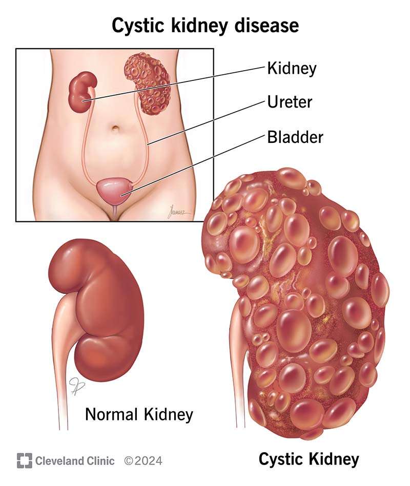
Kidney Diseases encompass a spectrum of conditions affecting kidney function, ranging from acute kidney injury (AKI) to chronic kidney disease (CKD). Effective management is crucial to prevent progression to end-stage renal disease (ESRD).
🩺 Treatment Approaches
1. Lifestyle Modifications
- Dietary Adjustments: Implementing a renal diet that limits sodium, phosphorus, potassium, and protein intake can alleviate kidney workload. This dietary approach helps manage electrolyte imbalances and reduces fluid retention .
- Hydration: Maintaining adequate fluid intake is essential, but in certain cases, fluid restrictions may be necessary to prevent fluid overload. Monitoring urine output and adjusting fluid intake accordingly is vital .
- Physical Activity: Engaging in regular exercise helps control blood pressure and improves overall health, which is beneficial for kidney function .
2. Pharmacological Interventions
- Blood Pressure Management: Angiotensin-converting enzyme (ACE) inhibitors and angiotensin II receptor blockers (ARBs) are commonly prescribed to control blood pressure and reduce proteinuria, thereby slowing CKD progression .
- Anemia Treatment: Erythropoiesis-stimulating agents (ESAs) and iron supplements are used to address anemia associated with CKD .
- Cholesterol Control: Statins may be recommended to manage elevated cholesterol levels, reducing cardiovascular risks in CKD patients .
3. Renal Replacement Therapy (RRT)
- Dialysis: Hemodialysis and peritoneal dialysis are employed when kidneys can no longer effectively filter waste products. Hemodialysis uses a machine to filter blood, while peritoneal dialysis uses the lining of the abdomen .
- Kidney Transplantation: A kidney transplant involves replacing a diseased kidney with a healthy one from a donor. This option is considered for eligible patients with ESRD .
4. Emerging Therapies
- Daprodustat: An oral medication approved for treating anemia due to CKD, offering an alternative to injectable ESAs .
Conclusion
Managing kidney disease requires a multifaceted approach, including lifestyle modifications, pharmacological treatments, and, when necessary, renal replacement therapies. Early detection and consistent management are key to preserving kidney function and improving patient outcomes. Regular monitoring and collaboration with healthcare providers are essential components of effective kidney disease management.
BREAST AND THYROID DISEASES
Breast Diseases
1. Breast Cancer
The most common malignancy among women globally, breast cancer originates in the breast cells, often presenting as lumps, changes in breast shape, or skin dimpling. Risk factors include age, family history, hormonal exposure, and lifestyle choices. Treatment modalities encompass surgery, chemotherapy, radiation therapy, and hormone therapy, tailored to cancer type and stage.
2. Benign Breast Conditions
These include fibroadenomas, cysts, and fibrocystic changes, which are non-cancerous but may cause discomfort or anxiety. Management often involves monitoring, aspiration of cysts, or surgical removal if necessary.
🩺 Thyroid Diseases
1. Hypothyroidism
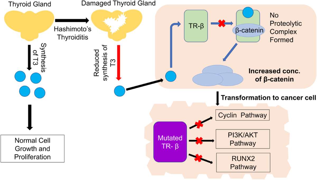
Characterized by insufficient thyroid hormone production, leading to symptoms like fatigue, weight gain, and depression. Common causes include Hashimoto's thyroiditis and iodine deficiency. Treatment typically involves thyroid hormone replacement therapy.
2. Hyperthyroidism
An overactive thyroid produces excess hormones, causing symptoms such as weight loss, rapid heartbeat, and anxiety. Graves' disease is a common cause. Management options include antithyroid medications, radioactive iodine therapy, or surgery.
3. Thyroid Cancer
Although rare, thyroid cancer can occur, with papillary carcinoma being the most common type. Treatment usually involves surgical removal of the thyroid gland, followed by radioactive iodine therapy.
🔗 Interconnections Between Breast and Thyroid Diseases
Recent studies suggest a potential link between thyroid disorders and an increased risk of breast cancer. For instance, hyperthyroidism and autoimmune thyroid diseases like Hashimoto's thyroiditis have been associated with a higher incidence of breast cancer. Conversely, hypothyroidism may be linked to a reduced risk, though findings are inconsistent and require further investigation.
Additionally, patients with thyroid cancer may have an elevated risk of developing breast cancer, particularly if they have a family history of cancer or have undergone radiation therapy.
Conclusion
Understanding the relationship between breast and thyroid diseases is crucial for early detection and management. Patients with thyroid disorders should be monitored for breast health, and vice versa, to ensure comprehensive care. Regular screenings and consultations with healthcare providers are recommended for individuals at risk.
HEMORRHOIDS
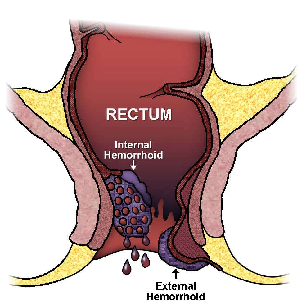
Hemorrhoids
Conservative Measures:
- Dietary Modifications: Increase fiber intake to 25–35 grams per day to soften stools and reduce straining.
- Hydration: Drink ample fluids to prevent constipation.
- Sitz Baths: Soak the anal area in warm water for 10–15 minutes several times daily to alleviate pain and swelling.
- Topical Treatments: Use over-the-counter creams or ointments containing hydrocortisone or witch hazel for symptom relief.
Medical Interventions:
- Rubber Band Ligation: A small rubber band is placed around the base of the hemorrhoid to cut off blood supply, causing it to shrink and fall off.
- Sclerotherapy: Injection of a chemical solution into the hemorrhoid to shrink it.
- Infrared Coagulation: Use of infrared light to cause scarring and shrinkage of the hemorrhoid.
Surgical Options:
- Hemorrhoidectomy: Surgical removal of large or prolapsed hemorrhoids.
- Stapled Hemorrhoidopexy: Stapling technique to remove hemorrhoidal tissue and reposition prolapsed hemorrhoids.
🩺 Anal Fissures Treatment
Conservative Measures:
- Fiber-Rich Diet: Consume 18–30 grams of fiber daily to prevent constipation.
- Hydration: Adequate fluid intake to soften stools.
- Sitz Baths: Warm water soaks to relax the anal sphincter and promote healing.
- Topical Analgesics: Use of lidocaine ointment for pain relief.
Medical Treatments:
- Nitroglycerin Ointment: Applied to the anal area to increase blood flow and reduce sphincter pressure, aiding healing.
- Botox Injections: Botulinum toxin injection to relax the anal sphincter muscle.
- Calcium Channel Blockers: Topical application of nifedipine or diltiazem to relax the anal sphincter.
Surgical Intervention:
- Lateral Internal Sphincterotomy (LIS): Surgical procedure involving a small incision in the anal sphincter to relieve pressure and promote healing.
Both conditions can often be managed with lifestyle modifications and medical treatments. However, persistent or severe cases may require surgical intervention. Consultation with a healthcare provider is recommended for proper diagnosis and treatment planning.
GASTRO-INTESTINAL DISEASE
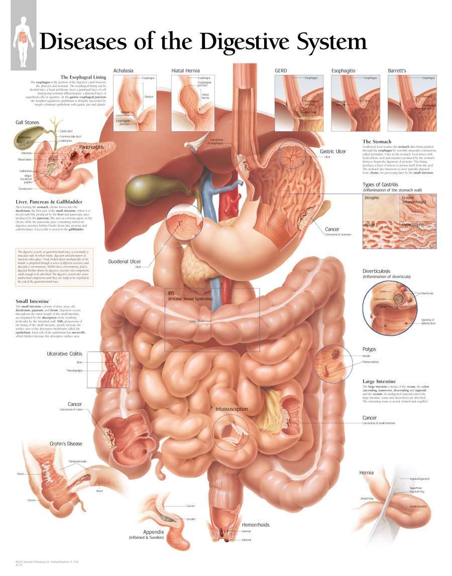
Gastrointestinal (GI) diseases encompass a wide range of conditions affecting the digestive system, including the esophagus, stomach, intestines, liver, and pancreas. These disorders can significantly impact daily life, causing symptoms such as abdominal pain, bloating, diarrhea, constipation, and nausea.
Common Gastrointestinal Disorders
- Gastroesophageal Reflux Disease (GERD): Characterized by chronic acid reflux, GERD leads to heartburn, chest pain, and regurgitation. It results from the backflow of stomach contents into the esophagus due to a weakened lower esophageal sphincter. Management includes lifestyle modifications, medications like proton pump inhibitors, and, in severe cases, surgery.
- Irritable Bowel Syndrome (IBS): A functional GI disorder causing symptoms like abdominal cramps, bloating, and alternating diarrhea and constipation. The exact cause remains unknown, but stress and diet can trigger symptoms. Treatment focuses on dietary changes, stress management, and medications to alleviate symptoms.
- Inflammatory Bowel Disease (IBD): Includes Crohn's disease and ulcerative colitis, conditions characterized by chronic inflammation of the gastrointestinal tract. Symptoms encompass persistent diarrhea, abdominal pain, fatigue, and weight loss. Treatment may involve anti-inflammatory drugs, immune system suppressors, and sometimes surgery.
- Peptic Ulcer Disease: Caused by the erosion of the stomach lining or the upper part of the small intestine, leading to sores or ulcers. Symptoms include stomach pain, bloating, and indigestion. Treatment typically involves antibiotics for H. pylori infection and medications to reduce stomach acid.
- Celiac Disease: An autoimmune disorder where ingestion of gluten damages the small intestine lining, leading to malabsorption of nutrients. Symptoms vary widely and can include diarrhea, weight loss, and fatigue. The primary treatment is a strict, lifelong gluten-free diet.
Diagnosis and Treatment
Diagnosis of GI diseases often involves a combination of medical history review, physical examination, laboratory tests, imaging studies, and endoscopic procedures. Treatment strategies are tailored to the specific condition and may include dietary modifications, medications, and in some cases, surgical interventions.
Conclusion
Gastrointestinal diseases are prevalent and can range from mild to severe. Early diagnosis and appropriate management are crucial for improving quality of life and preventing complications. If you experience persistent digestive symptoms, consult a healthcare professional for evaluation and personalized treatment.
HERNIA
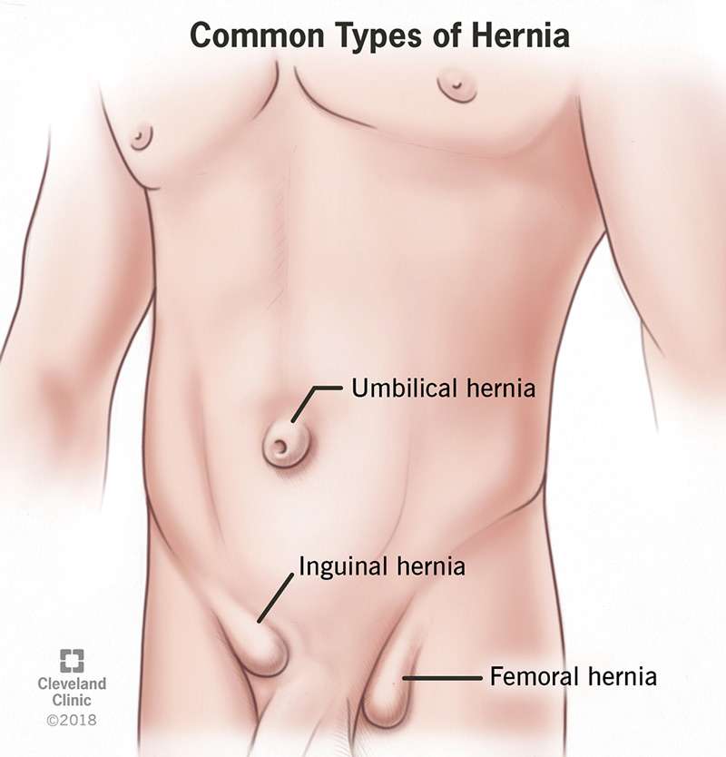
A Hernia occurs when an internal organ or tissue bulges through a weak spot in the abdominal wall, often causing pain or discomfort. While some small, asymptomatic hernias may be managed conservatively, surgical intervention is typically recommended for symptomatic or complicated cases.
Surgical Options
- Open Hernia Repair: This traditional method involves making a single incision at the hernia site to reposition the protruding tissue and reinforce the abdominal wall, usually with a synthetic mesh. It's effective for various hernia types but may require a longer recovery period.
- Laparoscopic (Minimally Invasive) Repair: Utilizing several small incisions, this technique involves inserting a camera and instruments to repair the hernia. It offers benefits like reduced pain, shorter hospital stays, and quicker recovery. Laparoscopic repair is often preferred for bilateral or recurrent hernias.
- Robotic-Assisted Surgery: An advanced form of laparoscopic surgery, robotic-assisted procedures provide enhanced precision and control. The surgeon operates robotic arms while viewing a 3D magnified image, improving outcomes, especially in complex or large hernias.
Conservative Management
For small, asymptomatic hernias, a "watchful waiting" approach may be advised. This involves monitoring the hernia for any changes or worsening symptoms over time. Lifestyle modifications, such as avoiding heavy lifting and maintaining a healthy weight, can help manage symptoms.
Conclusion
The choice of treatment depends on the hernia's type, size, and location and the patient's overall health. Consulting with a healthcare professional is essential to determine the most appropriate treatment plan.
UROLOGY
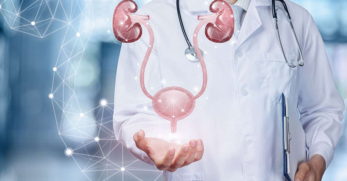
Urology is a medical specialty focusing on the diagnosis and treatment of conditions affecting the urinary tract and male reproductive organs. Urologists manage a wide range of disorders, including urinary tract infections (UTIs), kidney stones, benign prostatic hyperplasia (BPH), prostate cancer, erectile dysfunction, and incontinence.
Common Urological Treatments
- Medications: Alpha-blockers and 5-alpha-reductase inhibitors are commonly prescribed to manage BPH symptoms by relaxing prostate muscles and reducing gland size.
- Minimally Invasive Procedures:
- Transurethral Microwave Thermotherapy (TUMT): Utilizes microwave energy to shrink prostate tissue, alleviating BPH symptoms.
- Prostatic Artery Embolization (PAE): A non-surgical technique that blocks blood flow to the prostate, leading to its shrinkage.
- Surgical Interventions:
- Transurethral Resection of the Prostate (TURP): A standard procedure for BPH involving the removal of prostate tissue through the urethra.
- Prostatectomy: Surgical removal of all or part of the prostate gland, often performed for prostate cancer.
- Neuromodulation Therapy:
- Percutaneous Tibial Nerve Stimulation (PTNS): A minimally invasive procedure that stimulates nerves controlling the bladder, effective for overactive bladder symptoms.
GENERAL SURGERY
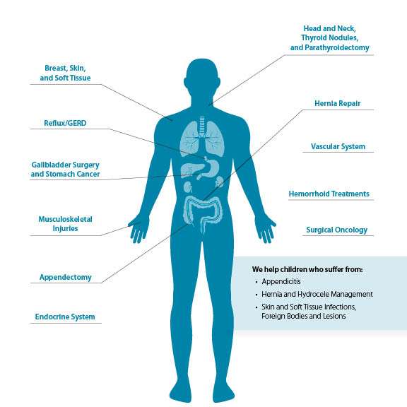
General surgery is a medical specialty that focuses on surgical treatment of conditions affecting the abdomen, digestive tract, skin, soft tissues, and endocrine system. It includes both elective and emergency procedures, often performed using advanced techniques such as laparoscopy (keyhole surgery) and robotic-assisted systems.
Common General Surgical Treatments:
- Appendectomy: Surgical removal of an inflamed appendix, commonly due to appendicitis. This is often an emergency procedure.
- Cholecystectomy: Removal of the gallbladder, typically performed to treat gallstones or gallbladder infection.
- Hernia Repair: Fixes abdominal wall hernias using sutures or surgical mesh, done through open or laparoscopic techniques.
- Bowel Resection: Removal of a diseased part of the intestine, often necessary for cancer, Crohn’s disease, or obstruction.
- Mastectomy: Surgical removal of one or both breasts, usually as a treatment for breast cancer.
- Thyroidectomy: Partial or total removal of the thyroid gland to treat thyroid disorders or cancer.
Modern Techniques:
Minimally invasive surgery, such as laparoscopic and robotic surgery, uses small incisions, reducing recovery time, pain, and hospital stay. These approaches are increasingly preferred due to lower risk of complications.
Postoperative Care:
Recovery varies by procedure but usually includes pain management, wound care, mobility exercises, and follow-up visits. Lifestyle changes and medications may be recommended to support long-term healing.
STONE DISEASES
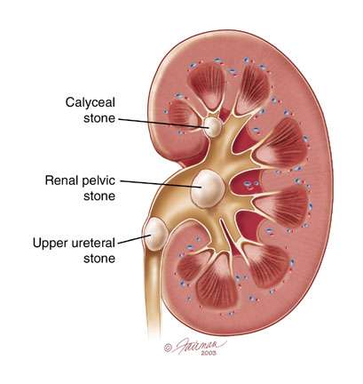
Stone diseases, particularly kidney stones, require treatment based on the type, size, and location of the stone, as well as the severity of symptoms. Small stones often pass naturally through the urinary tract with increased fluid intake. Drinking 2–3 liters of water daily helps dilute the urine, making it easier for small stones to pass. Pain can be managed with over-the-counter medications like ibuprofen or prescribed painkillers.
For larger or symptomatic stones, medical intervention may be necessary. Medications such as alpha-blockers (e.g., tamsulosin) can relax the muscles in the ureter, aiding stone passage. Uric acid stones may dissolve with urine alkalinization using potassium citrate or sodium bicarbonate.
VENTRICULAR ASSIST DEVICE (VAD) IMPLANTATION
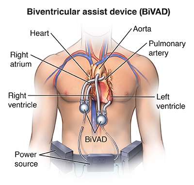
Ventricular Assist Device (VAD) implantation is a surgical procedure that involves the placement of a mechanical pump or device to assist the pumping function of a weakened or failing heart. VADs are primarily used as a bridge to heart transplantation or as destination therapy for patients who are not eligible for heart transplantation.
VAD implantation is considered for individuals with severe heart failure who have exhausted other treatment options and have a poor prognosis without intervention. A thorough evaluation is performed to assess the patient's overall health, heart function, and suitability for VAD therapy.
There are different types of VADs available, including:
- Left Ventricular Assist Device (LVAD): This device is implanted to assist the pumping function of the left ventricle, which is the main pumping chamber of the heart.
- Right Ventricular Assist Device (RVAD): In some cases, a separate device may be implanted to support the right ventricle's function.
- Biventricular Assist Device (BiVAD): This involves the implantation of both an LVAD and an RVAD to support both ventricles.
- Total Artificial Heart: In rare cases of severe biventricular failure, a total artificial heart may be implanted to replace both ventricles temporarily until a heart transplant becomes available.
The VAD implantation procedure is typically performed under general anesthesia. The surgeon makes an incision in the chest and places the device in the appropriate position, which can be in the abdomen or the chest cavity. Tubes are connected to the heart chambers to divert blood flow through the VAD. The VAD is then connected to an external power source and controller.
Following the surgery, the patient is closely monitored in the intensive care unit (ICU) to ensure proper functioning of the VAD, stability of the patient, and to manage any immediate complications. The patient may remain in the hospital for several weeks to receive specialized care and support during the recovery phase.
Once the patient is stable, a rehabilitation program may be initiated to promote physical recovery and help the patient regain strength. The patient and their caregivers also receive education and training on VAD management, including how to maintain and troubleshoot the device, manage medications, and recognize signs of potential complications.
After leaving the hospital, the patient continues to receive regular follow-up care from a specialized VAD team. This includes monitoring VAD function, managing medications, conducting routine tests, and addressing any issues or concerns that may arise.
Ventricular Assist Devices can significantly improve the quality of life and survival for individuals with advanced heart failure. While VADs can provide life-saving support, they are associated with certain risks and complications, such as bleeding, infection, device malfunction, blood clots, and complications related to the use of anticoagulant medications. Close monitoring and adherence to the VAD management plan are crucial for optimal outcomes.
The decision to undergo VAD implantation is made based on a careful assessment of the patient's specific condition, medical history, and overall health. It is a complex decision-making process that involves collaboration among the patient, the healthcare team, and the patient's family/support system.
HEART TRANSPLANTATION
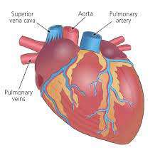
Heart transplantation is a surgical procedure in which a diseased or failing heart is replaced with a healthy heart from a deceased donor. It is performed on individuals with end-stage heart failure or severe heart conditions that cannot be effectively treated with other therapies.
Before a heart transplant, a comprehensive evaluation is conducted to assess the patient's overall health, heart function, and suitability for transplantation. This evaluation includes medical tests, imaging studies, blood work, and psychosocial assessments. If the patient meets the criteria, they are placed on a waiting list for a suitable donor heart.
When a suitable donor heart becomes available, a series of tests are performed to ensure compatibility and minimize the risk of rejection. The donor heart is transported to the recipient's hospital, and the surgical team prepares for the transplantation procedure. The recipient is placed under general anesthesia.
The surgical team makes an incision in the chest to access the heart. The recipient's diseased heart is removed, and the donor heart is carefully implanted and connected to the recipient's blood vessels, including the aorta and pulmonary artery. The new heart is then reconnected to the recipient's remaining heart structures, such as the remaining atria.
Once the new heart is in place, blood flow is restored to the transplanted heart, and the team ensures that the heart is functioning properly. The surgical team monitors the patient closely during this critical phase to ensure that the new heart is effectively pumping blood and that the recipient is stable.
After the surgery, the patient is transferred to the intensive care unit (ICU) for close monitoring. The recovery process involves a multidisciplinary team of healthcare professionals who provide post-transplant care, including monitoring for signs of organ rejection, managing immunosuppressive medications to prevent rejection, and addressing any complications that may arise.
Following the initial recovery period, the patient is transferred to a regular hospital room and eventually discharged. Rehabilitation plays a vital role in the patient's recovery, including physical therapy, cardiac rehabilitation, and lifestyle modifications to support heart health. Regular follow-up visits with the transplant team are essential to monitor the patient's heart function, manage medications, and address any issues or concerns.
Heart transplantation is a complex and life-saving procedure, but it is not without risks. The major risks include rejection of the transplanted heart, infection, complications from immunosuppressive medications, and the potential need for future transplant-related procedures.
However, heart transplantation has shown to significantly improve survival rates and quality of life for individuals with end-stage heart failure. The decision to undergo a heart transplant is made based on careful consideration of the patient's specific condition, medical history, and overall health, and it requires a collaborative approach involving the patient, the transplant team, and other healthcare professionals.
LUNG SURGERY
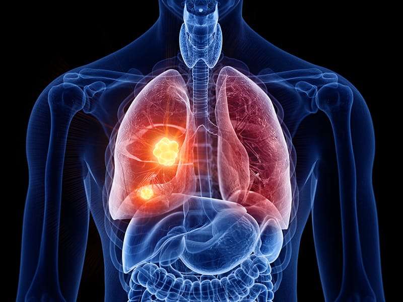
Lung surgery, also known as thoracic surgery, refers to surgical procedures performed on the lungs or other structures within the thoracic cavity. Lung surgery is often used to diagnose and treat various lung conditions, including lung cancer, lung infections, lung nodules, and certain lung diseases.
Lung surgery may be recommended for several reasons, including the removal of lung tumors (benign or malignant), treatment of lung infections (such as abscesses or empyema), management of lung conditions like bullae or lung cysts, and treatment of certain lung diseases like lung volume reduction surgery for advanced emphysema.
Prior to lung surgery, a thorough evaluation is conducted to assess the patient's overall health, lung function, and the extent of the lung condition. This evaluation may involve imaging studies (such as CT scans or PET scans), pulmonary function tests, blood work, and biopsies to determine the precise nature of any lung masses or nodules.
The surgical approach and incision depend on the specific procedure being performed. Traditional open surgery involves making an incision in the chest wall, often in the area between the ribs. Minimally invasive techniques, such as video-assisted thoracoscopic surgery (VATS), may be used, which involve smaller incisions and the use of specialized instruments and a video camera to access and visualize the lungs.
The specific surgical procedure depends on the underlying lung condition. Common lung surgeries include:
- Lobectomy: Removal of an entire lobe of the lung. This is often performed for lung cancer or certain benign lung conditions.
- Wedge Resection: Removal of a small, wedge-shaped portion of lung tissue. This may be performed for localized lung tumors or to remove lung nodules.
- Pneumonectomy: Removal of an entire lung. This is typically reserved for extensive lung tumors or in cases where one lung is severely damaged or infected.
- Segmentectomy: Removal of a specific segment of the lung. This procedure is performed when removing an entire lobe is not necessary, typically for smaller lung tumors or localized lung disease.
- Pleurodesis: A procedure to treat recurrent pleural effusion (fluid accumulation between the lungs and chest wall). It involves creating adhesions between the lung and chest wall to prevent fluid buildup.
Following lung surgery, patients typically require regular follow-up visits with their healthcare team. Rehabilitation and respiratory therapy may be recommended to aid in recovery and improve lung function. This may involve breathing exercises, physical activity, and guidance on lifestyle modifications to promote lung health.
It is important to note that lung surgery is a complex procedure and carries potential risks and complications, including bleeding, infection, blood clots, respiratory issues, and reactions to anesthesia. The specific approach and procedure chosen depend on the patient's condition, overall health, and the expertise of the surgical team. The decision to undergo lung surgery is made on an individual basis, weighing the potential benefits against the risks, and considering other
HEART VALVE REPAIR OR REPLACEMENT
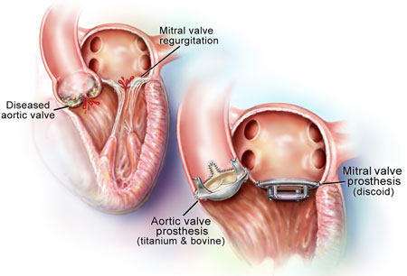
Heart valve repair or replacement is a surgical procedure performed to treat heart valve diseases that involve damage or dysfunction of one or more of the heart's valves. The procedure aims to restore proper valve function and improve overall heart function.
Here's an overview of heart valve repair or replacement:
Heart valve repair or replacement may be recommended for various conditions, including valvular stenosis (narrowing of the valve) or regurgitation (leakage of blood backward through the valve). Common valve conditions that may require intervention include aortic valve stenosis or regurgitation, mitral valve regurgitation or stenosis, tricuspid valve regurgitation, and pulmonic valve stenosis or regurgitation.
Before the procedure, a comprehensive evaluation is conducted, which may include echocardiography, cardiac catheterization, stress tests, and other imaging studies to assess the valve's structure and function, determine the severity of the disease, and evaluate overall heart function.
The surgeon evaluates the valve and determines whether repair or replacement is necessary. Valve repair involves techniques to preserve and restore the natural valve structure, such as removing excess tissue, repairing the valve leaflets, or reinforcing the valve with a prosthetic ring. Valve replacement involves removing the damaged valve and replacing it with a mechanical valve (made of synthetic materials) or a biological valve (made from human or animal tissue).
After repairing or replacing the valve, the surgeon tests the valve's function and closes the incisions. Temporary pacing wires may be left in place to monitor the heart's electrical activity during the recovery period.
Following the procedure, the patient is transferred to the intensive care unit (ICU) for close monitoring. Over the next few days, the patient gradually transitions to a regular hospital room. Physical therapy, breathing exercises, and medications are provided to promote healing and recovery.
The patient may need to take blood-thinning medications (in the case of mechanical valve replacement) or other medications to prevent infection or manage symptoms. Heart valve repair or replacement is a major surgical procedure, and like any surgery, it carries risks such as bleeding, infection, blood clots, or complications associated with anesthesia.
CORONARY ARTERY BYPASS GRAFTING (CABG)
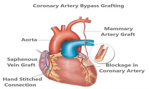
Coronary Artery Bypass Grafting (CABG) is a surgical procedure used to treat coronary artery disease (CAD), a condition characterized by the narrowing or blockage of the coronary arteries that supply blood to the heart muscle. CABG aims to restore normal blood flow to the heart by bypassing the blocked or narrowed arteries using grafts from other blood vessels.
Here's an overview of the CABG procedure:
1. Indications: CABG is typically recommended for individuals with severe coronary artery disease who experience symptoms such as chest pain (angina) or those who are at high risk of a heart attack. It is also considered for individuals with complex coronary artery blockages or when other treatments, such as medications or angioplasty, are not sufficient.
2. Preoperative Evaluation: Before undergoing CABG, a thorough evaluation is performed to assess the extent of coronary artery disease, heart function, and overall health. This evaluation may include tests such as angiography, stress tests, echocardiography, and blood work.
3. Anesthesia: CABG is performed under general anesthesia, meaning the patient is unconscious during the procedure. The anesthesiologist closely monitors vital signs and administers medications to ensure comfort and safety.
4. Incision and Exposure: The surgeon makes an incision in the middle of the chest (median sternotomy) to access the heart. Alternatively, minimally invasive techniques may be used, such as small incisions between the ribs (minithoracotomy) or robotic-assisted surgery.
5. Harvesting Grafts: Blood vessels are harvested from other parts of the body, typically the internal mammary artery from the chest wall and/or veins (saphenous veins) from the leg. These grafts will be used to bypass the blocked or narrowed coronary arteries.
6. Cardiopulmonary Bypass: To allow the surgeon to work on the heart, a heart-lung bypass machine is connected to the patient. The machine takes over the function of the heart and lungs, circulating and oxygenating blood throughout the body while the surgeon performs the bypass procedure.
7. Grafting: The surgeon carefully attaches the grafts to the coronary arteries, bypassing the blockages. The grafts serve as new pathways for blood to flow to the heart muscle, bypassing the diseased segments of the coronary arteries.
8. Conclusion: After completing the grafting, the surgeon verifies the grafts' function and closes the incisions. Temporary pacing wires may be left in place to monitor the heart's electrical activity during the recovery period.
9. Recovery: Following the procedure, the patient is transferred to the intensive care unit (ICU) for close monitoring. Over the next few days, the patient gradually transitions to a regular hospital room. Physical therapy, breathing exercises, and medications are provided to promote healing and recovery.
CABG is a major surgical procedure and, like any surgery, carries risks such as bleeding, infection, blood clots, and complications associated with anesthesia. However, it is a well-established procedure with a high success rate in relieving angina symptoms, improving quality of life, and prolonging survival in appropriate patients with severe coronary artery disease.
INTERVENTIONAL PROCEDURES

Interventional procedures are medical procedures that are performed using minimally invasive techniques to diagnose or treat various conditions. These procedures often involve the use of specialized instruments, imaging guidance, and small incisions or catheter insertions. They are typically performed by interventional radiologists, cardiologists, or other specialized physicians. Common interventional procedures include angiography and angioplasty, where angiography involves injecting a contrast dye into blood vessels to visualize their structure and identify abnormalities, while angioplasty is used to widen narrowed or blocked blood vessels using a balloon catheter to restore blood flow. Stent placement is another commonly performed procedure in which small mesh tubes are inserted into narrowed or blocked blood vessels to keep them open, particularly in patients with coronary artery disease, peripheral artery disease, and other vascular conditions. Biopsy procedures involve removing a small sample of tissue for examination and diagnosis, often using imaging guidance such as ultrasound or CT scans for precise targeting. Radiofrequency ablation utilizes heat generated by radiofrequency energy to destroy abnormal or cancerous cells and is frequently used for tumors of the liver, kidney, lung, or bone. Embolization is a technique used to block or reduce blood flow to specific areas by injecting embolic agents into blood vessels, making it effective for treating bleeding, tumors, and vascular malformations. Vertebroplasty and kyphoplasty are specialized procedures designed to stabilize fractured vertebrae by injecting bone cement into the affected spinal bones, thereby providing support and pain relief. Chemoembolization and radioembolization deliver chemotherapy drugs or radioactive particles directly into tumors or the blood vessels supplying them, offering targeted treatment for liver cancer and certain metastatic tumors. Drainage procedures are performed to remove fluid or pus from body cavities or abscesses, often with the assistance of image-guided catheter placement. Additionally, ablation procedures for arrhythmias, including radiofrequency ablation and cryoablation, are used to treat abnormal heart rhythms by selectively destroying abnormal electrical pathways within the heart.
CARDIOLOGY

PSYCHIATRY

RHEUMATOLOGY
VASCULAR
PEDIATRICIAN & NICU

SURGERY & TRAUMA CARE
ORTHOPAEDIC
CARDIOLOGY